HUMAN LOCOMOTION SYSTEM презентация
Содержание
- 2. Human Skeletal System Functions: Supports the body Provides movement with
- 3. Skeletal System Skeletal system is the supportive and protective system of
- 4. Structure of human skeleton Human skeleton is composed of bones and
- 7. Bone Outer cover of bones known as periosteum Periosteum provides growth
- 9. Red and Yellow bone marrows
- 10. Formation of bone For bone formation and normal growth minerals such
- 12. Types of bones 1. Long bones: Ex: bones of legs, arms
- 13. Parts of Human Skeletal System Adult human contains 206 bones, in
- 15. 1. Skull Skull includes 22 bones 8 of them cranial,
- 17. 2. Trunk Trunk includes vertebral column, ribs, sternum, pelvic girdle and
- 19. Chest Chest It protects heart and lungs that has 12 pairs
- 20. Pectoral girdle – плечевой пояс It includes 2 paired bones that
- 21. Pelvic girdle – тазовый пояс It is connected to the lower
- 22. Vertebral column is divided into 5 parts Vertebral column is divided
- 25. 3. Extremites - конечности Extremites in other words appendages include upper
- 26. b. Lower extremites: b. Lower extremites: 2 legs, include 30 bones
- 28. Joints Joint forms the junction between two or more bones There
- 29. Types of joints
- 30. 1. Immovable joint Jointed bones cannot move All cranial and facial
- 31. 2. Slightly movable joints Connected by cartilage or connective tissue Vertebrae
- 32. 3. Movable joints The bones in movable joints are connected to
- 33. Types of movable joints Ball-and-socket Hinge Pivot
- 34. Joints
- 35. Types of joints
- 36. Disorders and diseases of human skeletal system Fractures – is a
- 37. Types of fractures
- 38. Osteoporosis
- 39. Rheumatoid arthritis
- 40. Muscular system Muscular system helps in the movement of body, inner
- 41. Human muscular system
- 43. Types of muscular tissue There are 3 types of muscular tissue,
- 45. 1. Smooth muscle Each cell is long, sharp-ended with a single
- 47. 2.Skeletal or striated muscle Cells are long, cylindrical and multinuclear, i.e.
- 49. Skeletal muscles cover the skeleton Skeletal muscles cover the skeleton They
- 51. 3.Cardiac muscle Cells are long, cylindrical, branched and with 1 nucleus
- 53. Structure of muscles Skeletal muscle tissues are composed of bundle of
- 54. Skeletal muscle structure
- 55. Contraction of muscles Actin and myozin proteins slide on each other
- 56. Muscle contraction
- 57. Interaction of muscles and skeleton Most skeletal muscles are attached to
- 58. A flexor muscle causes a joint to bend. An extensor muscle
- 59. Locomotion system outlines: 1. Skeletal system Functions Structure Bone formation,
- 60. Locomotion system objectives Explain the general function of skeletal system.
- 61. Locomotion system key terms
- 62. Скачать презентацию


Слайды и текст этой презентации
Слайд 1
Описание слайда:
HUMAN LOCOMOTION SYSTEM
Слайд 2
Описание слайда:
Human Skeletal System
Functions:
Supports the body
Provides movement with the help of muscles
Protects inner organs
Produces blood cells
Stores minerals such as P (phosphorus) and Ca (calcium)
Слайд 3
Описание слайда:
Skeletal System
Skeletal system is the supportive and protective system of organisms
There 2 types of skeletal system in organisms:
Exoskeleton: seen mainly in invertebrates
Endoskeleton: seen in vertebrates, sea stars, sponges
Слайд 4
Описание слайда:
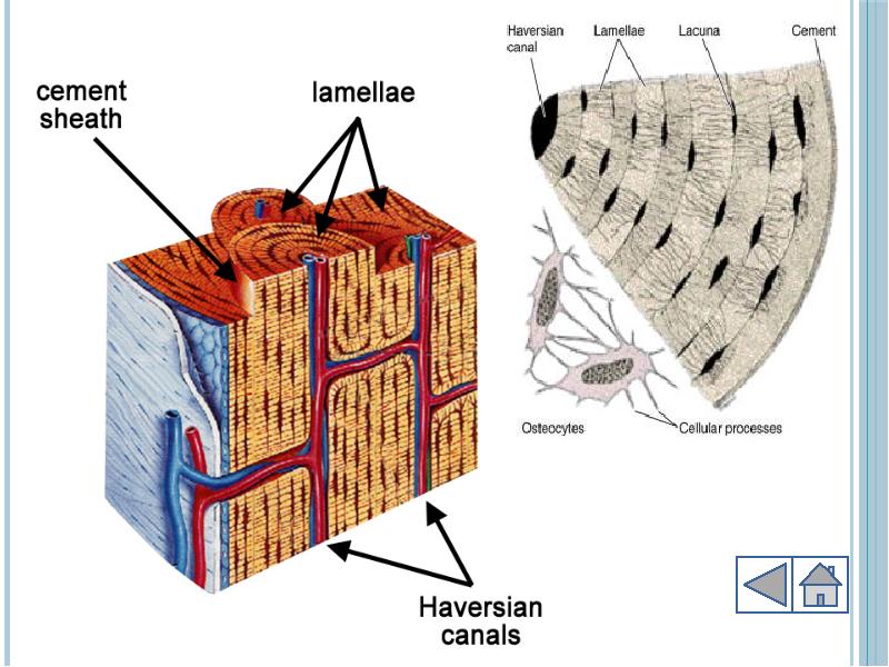
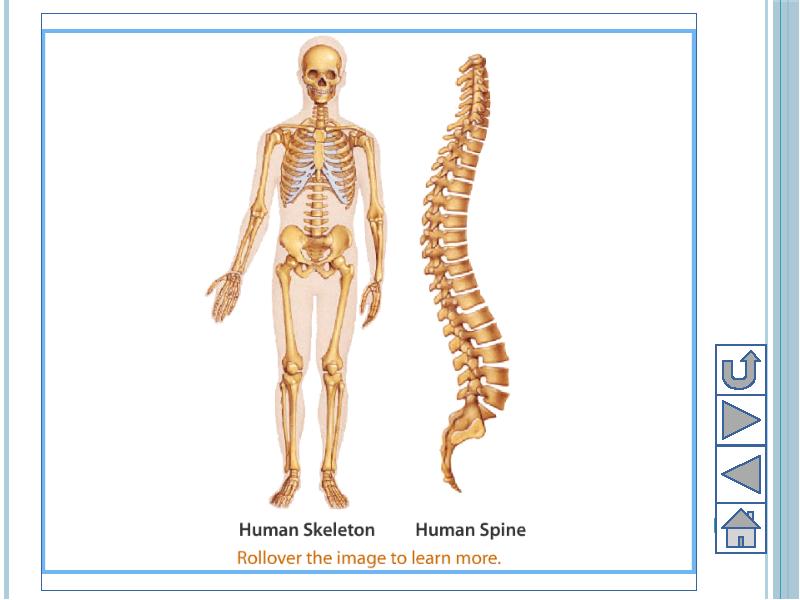
Structure of human skeleton
Human skeleton is composed of bones and cartilage
Bones are composed of cells known as osteocytes
Osteocytes are arranged in circles and connected to each other by cytoplasmic bridges
There is haversian canal between circles, it contains blood vessels and nerves
Intracellular space is filled by matrix (ossein) that contain Ca, P, carbonate and protein.
Cartilage is composed of cells chondrocytes
Слайд 5
Описание слайда:
Слайд 6
Описание слайда:
Слайд 7
Описание слайда:
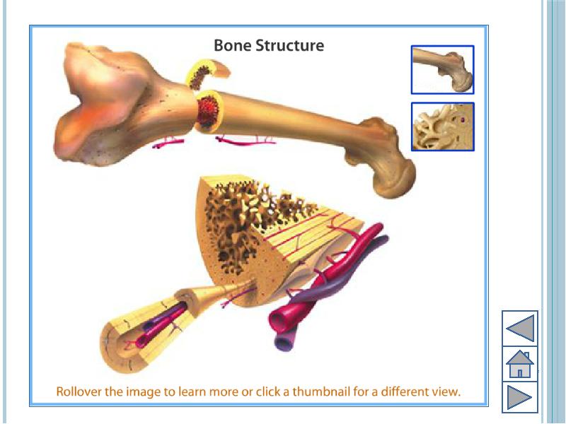
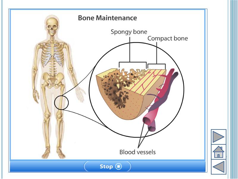
Bone
Outer cover of bones known as periosteum
Periosteum provides growth in diameter and repair of bones
There are 2 types of bone tissue:
Compact: very dense and strong
Spongy: porous and soft
Space between bones is filled with bone marrow
Red bone marrow: fills space between spongy bones and produces blood cells
Yellow bone marrow: fills hollow interior space of bones
Слайд 8
Описание слайда:
Слайд 9
Описание слайда:
Red and Yellow bone marrows
Слайд 10
Описание слайда:
Formation of bone
For bone formation and normal growth minerals such as Ca, P and vitamins A, C and D needed
Deficiency of vitamin D causes rickets – рахит, A growth rate decreases, C causes weakness and disease scurvy – цинга
Ca level in blood is regulated by hormones parathormone, released by parathyroid gland, and calcitonin, released by thyroid gland
Parathormone: is secreted when Ca level is decreased in blood
Calcitonin: is secreted when Ca level is increased from blood
Слайд 11
Описание слайда:
Слайд 12
Описание слайда:
Types of bones
1. Long bones:
Ex: bones of legs, arms …
2. Flat bones:
Ex: bones of skull, rib, patella …
3. Short bones:
Ex” bones of vertebrae, hand, fingers, foot …
Слайд 13
Описание слайда:
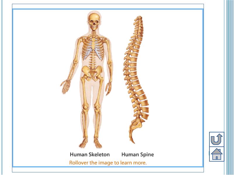
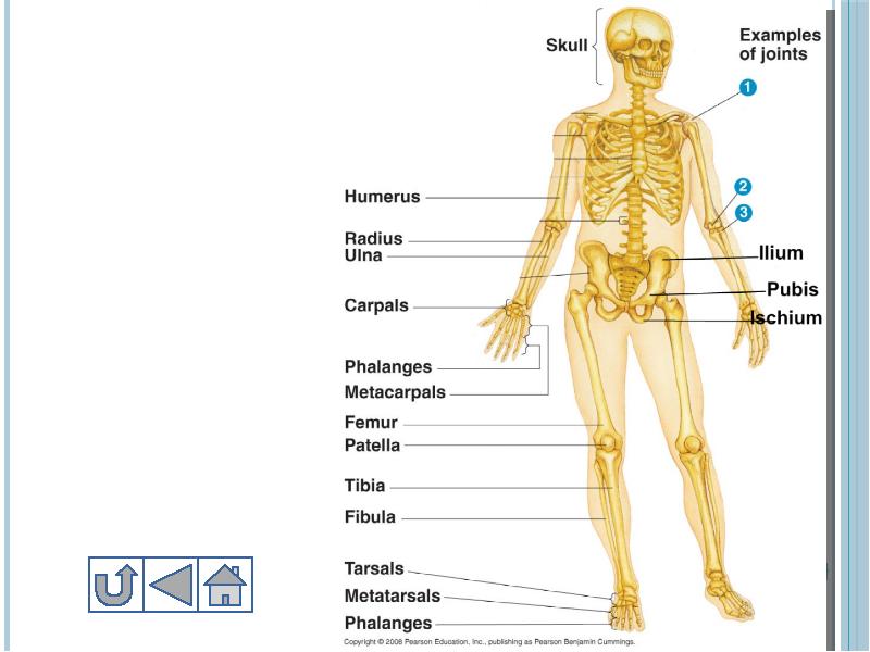
Parts of Human Skeletal System
Adult human contains 206 bones, in babies it is approximately 300
Skeleton parts:
Skull
Trunk
Extremites
Слайд 14
Описание слайда:
Слайд 15
Описание слайда:
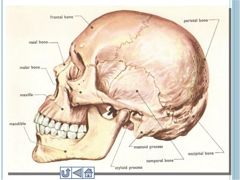
1. Skull
Skull includes 22 bones
8 of them cranial, 14 are facial bones
Cranial bones are fused to each other and immovable
Слайд 16
Описание слайда:
Слайд 17
Описание слайда:
2. Trunk
Trunk includes vertebral column, ribs, sternum, pelvic girdle and pectoral girdle
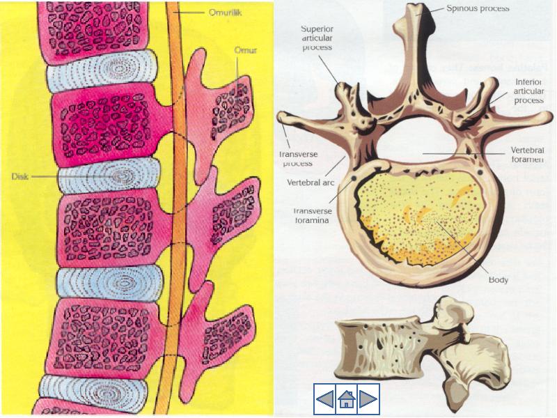
Vertebral column:
Vertebral column consists of 33 vertebrae
Between each vertebrae there is cartilaginous disc, and vertebral column is slightly movable
Vertebral column protects spinal cord
Upper end connected to Skull, lower end to Sacrum
Слайд 18
Описание слайда:
Слайд 19
Описание слайда:
Chest
Chest
It protects heart and lungs that has 12 pairs of ribs and a sternum.
All ribs are connected to vertebrae on the back side.
Ribs are connected to sternum in the abdomen. But 11th and 12th ribs are free floating ribs.
Слайд 20
Описание слайда:
Pectoral girdle – плечевой пояс
It includes 2 paired bones that hold up arms:
Clavicle
Scapula
Слайд 21
Описание слайда:
Pelvic girdle – тазовый пояс
It is connected to the lower end of vertebral column (sacrum)
It holds legs and reproductive organs
Includes:
Ilium
Ischium
Pubis
Слайд 22
Описание слайда:
Vertebral column is divided into 5 parts
Vertebral column is divided into 5 parts
1. Cervical or neck – 7 vertebrae
2. Thoracic – 12 vertebrae
3. Lumbar – 5 vertebrae (largest)
4. Sacral – 5 fused vertebrae
5. Coccyx or tailbone – 3 to 5 fused vertebrae
Слайд 23
Описание слайда:
Слайд 24
Описание слайда:
Слайд 25
Описание слайда:
3. Extremites - конечности
Extremites in other words appendages include upper extremites and lower extremites
a. Upper:
2 arms and include 30 bones each, bones:
Humerus 1*2
Ulna 1*2
Radius 1*2
Carpals 8*2
Metacarpals 5*2
Phalanges 14*2
Слайд 26
Описание слайда:
b. Lower extremites:
b. Lower extremites:
2 legs, include 30 bones each, bones:
Femur 1*2
Patella 1*2
Tibia 1*2
Fibula 1*2
Tarsals 7*2
Metatarsals 5*2
Phalanges 14*2
Слайд 27
Описание слайда:
Слайд 28
Описание слайда:
Joints
Joint forms the junction between two or more bones
There are three types of joints;
Immovable joints
Slightly movable joints
Movable joints
Слайд 29
Описание слайда:
Types of joints
Слайд 30
Описание слайда:
1. Immovable joint
Jointed bones cannot move
All cranial and facial bones except mandible and sacrum are immovable
There is no synovial fluid between bones
Слайд 31
Описание слайда:
2. Slightly movable joints
Connected by cartilage or connective tissue
Vertebrae are connected by cartilage and ribs to sternum also connected by cartilage
Слайд 32
Описание слайда:
3. Movable joints
The bones in movable joints are connected to each other by ligaments (very strong connective tissue fibers).
There is a synovial fluid and cartilage in movable joints which reduce friction.
Слайд 33
Описание слайда:
Types of movable joints
Ball-and-socket
Hinge
Pivot
Слайд 34
Описание слайда:
Joints
Слайд 35
Описание слайда:
Types of joints
Слайд 36
Описание слайда:
Disorders and diseases of human skeletal system
Fractures – is a broken bone because of high physical impact or some other bone disorders
Osteoporosis – disease which occurs when there is not enough deposition of calcium in bones and because of that bones get weaker
Rheumatoid arthritis – occurs when cells of immune system attack the tissues around joints causing the tissue become inflamed
Слайд 37
Описание слайда:
Types of fractures
Слайд 38
Описание слайда:
Osteoporosis
Слайд 39
Описание слайда:
Rheumatoid arthritis
Слайд 40
Описание слайда:
Muscular system
Muscular system helps in the movement of body, inner organs and also helps in protection of body organs
The units of muscular system are cells known as myofibrils
Myofibrils have the ability to contract and relax
Слайд 41
Описание слайда:
Human muscular system
Слайд 42
Описание слайда:
Слайд 43
Описание слайда:
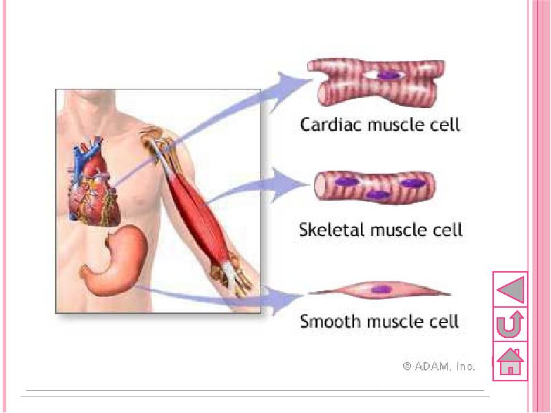
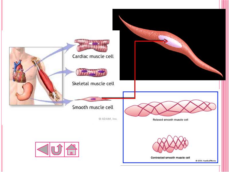
Types of muscular tissue
There are 3 types of muscular tissue, they are:
Smooth muscle
Skeletal muscle
Cardiac muscle
Слайд 44
Описание слайда:
Слайд 45
Описание слайда:
1. Smooth muscle
Each cell is long, sharp-ended with a single central nucleus
Smooth muscles generally regulated by the Autonomic Nervous System (ANS), i.e. we cannot control them (!)
They function involuntarily and movement is generally irregular and slow
They are found in the walls of inner organs, like stomach, intestine, blood vessels, urinary bladder etc.
Слайд 46
Описание слайда:
Слайд 47
Описание слайда:
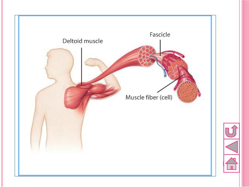
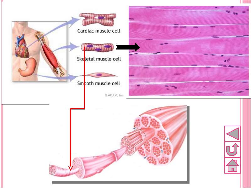
2.Skeletal or striated muscle
Cells are long, cylindrical and multinuclear, i.e. have many nucleuses
They are also termed as muscle fibers, because they are not branched
The structure of skeletal muscles:
Muscle bundles, muscle fibers, myofilaments (actin and myozin proteins)
Слайд 48
Описание слайда:
Слайд 49
Описание слайда:
Skeletal muscles cover the skeleton
Skeletal muscles cover the skeleton
They provide movement of skeleton, and mainly they are antagonistic
It is controlled by Somatic Nervous System (SNS)
It contracts rapidly
When it is overworked, maximal potential power is used, it gets hardened and this state is called as tetanus (судороги)
Слайд 50
Описание слайда:
Слайд 51
Описание слайда:
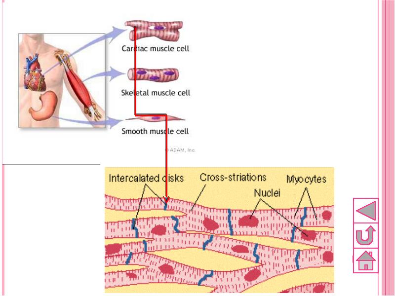
3.Cardiac muscle
Cells are long, cylindrical, branched and with 1 nucleus in the center of the cell
They have more mitochondria than skeletal muscles
Each cell is rich in blood and lymph vessels
As skeletal muscles they are rapid and because of its significant structure tetanus is not seen
They are controlled by ANS and function involuntarily
Слайд 52
Описание слайда:
Слайд 53
Описание слайда:
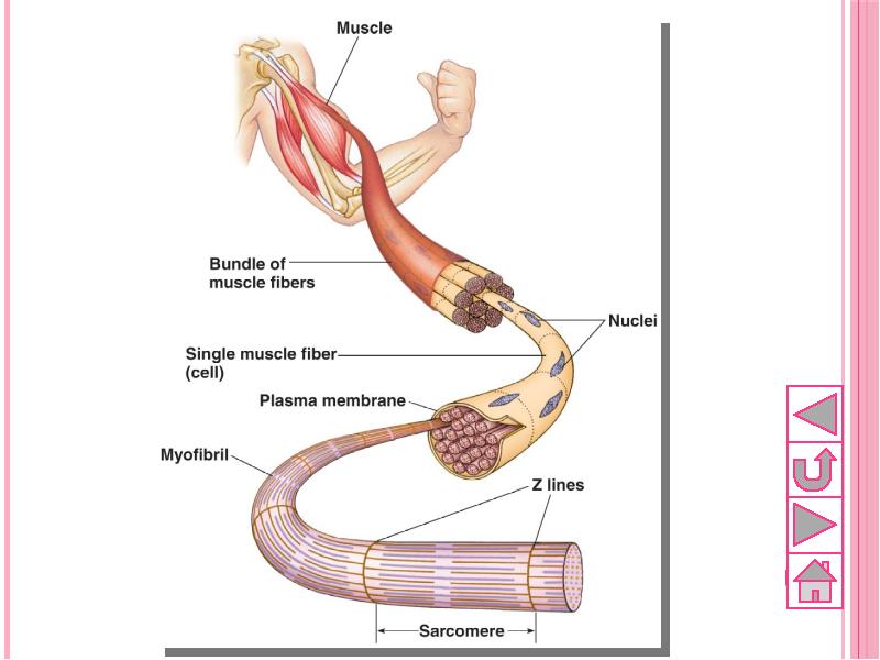
Structure of muscles
Skeletal muscle tissues are composed of bundle of muscle fibers, and muscle fibers are bundles of myofibrils
Myofibrils are divided into sarcomeres, the contraction units
Sarcomere contracts and relaxes by means of actin and myozin proteins
Слайд 54
Описание слайда:
Skeletal muscle structure
Слайд 55
Описание слайда:
Contraction of muscles
Actin and myozin proteins slide on each other and sarcomere shorten
For muscle contraction big amounts of energy and Ca2+ ions are needed
During contraction, CREATIN PHOSPHATE (which supplies 20 times more energy than ATP) is used as the primary energy source, ATP is used as the secondary.
During relaxation only ATP is used
Слайд 56
Описание слайда:
Muscle contraction
Слайд 57
Описание слайда:
Interaction of muscles and skeleton
Most skeletal muscles are attached to bones by strips of dense connective tissue called tendons.
Bones are connected each other at joints by means of fibrous connective tissue – ligaments.
Слайд 58
Описание слайда:
A flexor muscle causes a joint to bend. An extensor muscle causes a joint to straighten.
A flexor muscle causes a joint to bend. An extensor muscle causes a joint to straighten.
Слайд 59
Описание слайда:
Locomotion system
outlines:
1. Skeletal system
Functions
Structure
Bone formation, growth and types
Parts of human skeleton
Joints
Disorders and diseases of skeletal system
2. Muscular system
Functions
Types of muscle tissue
Structure of muscles
Muscle contraction
Interaction of muscles and skeleton
Слайд 60
Описание слайда:
Locomotion system
objectives
Explain the general function of skeletal system.
List the bones according to their shape
Describe the general structure of bones, and list the functions of its parts.
Distinguish between the axial and appendicular skeletons, and name the major parts of each.
Locate and identify the bones and major features of the bones that comprise the skull, vertebral column, rib cage, pectoral girdle, upper limb, pelvic girdle, lower limb.
List the three classes of joints describe their characteristics, and name an example of each.
Explain how skeletal muscles produce movements at joints, and identify several types of such movements.
Explain some metabolic and endocrine diseases of bones and skeletal system.
Name the major parts of a skeletal muscle fiber, and describe the function of each.
Explain the contraction mechanism of skeletal muscle.
Explain how the muscle fiber contraction mechanism obtains energy.
Describe how oxygen dept develops and how a muscle may become fatigued.
Compare the structure of skeletal, smooth and cardiac muscles.
Explain some inflammatory diseases of muscles.
Understand how muscle system helps other body systems and the relation between muscle and other body systems
Слайд 61
Описание слайда:
Locomotion system
key terms
Скачать презентацию на тему HUMAN LOCOMOTION SYSTEM можно ниже: