TOXOPLASMOSIS ( T.) Identification. Zoonotic disease caused by an о bligate презентация
Содержание
- 2. Historic reference. 1908 J.Nicolas and L. Manseaux in
- 3. The actuality of illness is determined by the following factors:
- 4. - absence for the doctors of common practice sufficient knowledge and
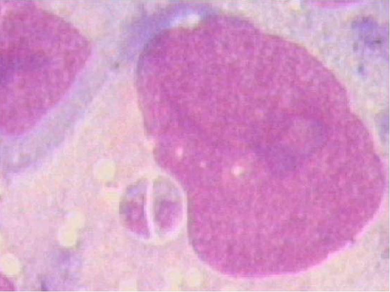
- 5. It is size from 2- 4 microns of width and to
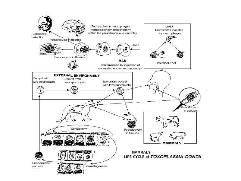
- 7. The biotic cycle Т. consists from sexual and asexual reproduction
- 8. Mature trophozoite is divided on merozoites, which part again penetrate into
- 9. Oocyst together with feces of the cat gets in the external
- 11. The main intermediate hosts - rats and mice, which some time
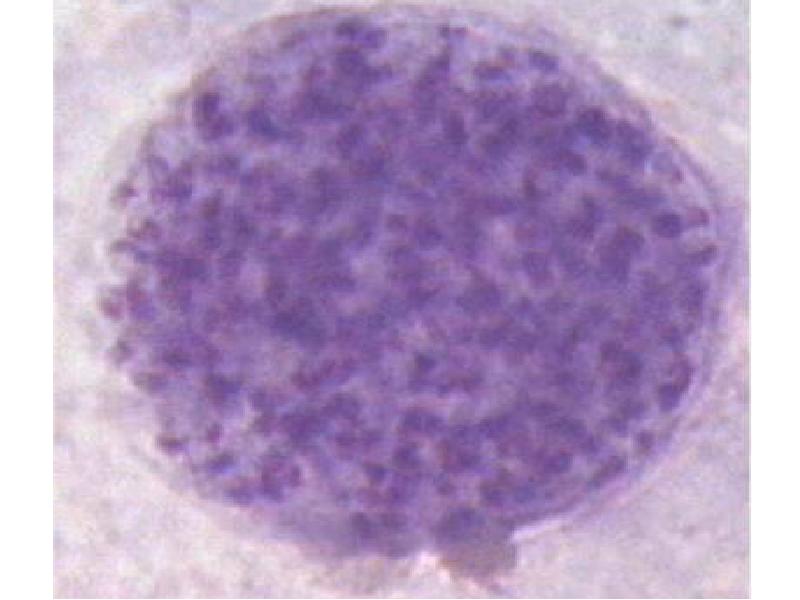
- 12. A nuclear cell before destroy ( “false cyst”)
- 13. After destroy a cell Т. penetrate into other cell recycling developments.
- 15. PATHOMORPHOLOGY The pathological changes are found out in all bodies both
- 16. It is possible to detect granulomas, centers of a necrosis and
- 17. EPIDEMIOLOGY (zoonosis with peroral and vertical mode of infection)
- 18. CLINIC (incubation interval about 2 weeks) Distinguish 2 forms Т.
- 21. To frequency there is a combination of numbered mani-festations by an
- 22. Objective: - generalized lymphadenopathy - increase of a liver (50 %)
- 23. - eyes - chorioretinitis (1 %), uveitis, progressing myopia -
- 24. CONGENITAL Т. - arises only at primary infection by the pregnant
- 25. Signs of disease is possible to detect for child at once
- 26. But it can pass in the chronic or secondary - latent
- 27. DIFFERENTIAL DIAGNOSTICS will be carried out by : - Catscratch disease
- 28. LABORATORY DIAGNOSIS. 1.Microscopy smears CSF ( at congenital Т.) – xanthochromatic,
- 29. TREATMENT The antiparasitic therapy Will not be carried out:
- 30. for strengthen operation of pyrimethaminum: - clindamycin 300 mg
- 31. For the pregnant woman at immunological peaking without clinical manifestations -
- 32. PROPHYLAXIS The special attention accesses on group of hazard among the
- 34. Скачать презентацию
 Identification. Zoonotic disease caused by an оbligate endocellular")


")


")

Distinguish 2 forms Т.")

")
, uveitis, progressing myopia
-")


")


Слайды и текст этой презентации
Слайд 1
Описание слайда:
TOXOPLASMOSIS ( T.) Identification. Zoonotic disease caused by an оbligate endocellular parasite Toxoplasma gondii, characteristic by chronic current, lymphadeno-pathy, increase of a liver and spleen, damage
of the nervous system, cross-striated muscles, myocardium, eyes and often infection of a fetus in acute period of illness for the pregnant woman
Слайд 2
Описание слайда:
Historic reference.
1908 J.Nicolas and L. Manseaux in Tunis detected of the infectious agent for a local rodent Ctenodactylus gondii.
The infectious agent has received name under of the form
body (toxon - arc, plasma - form)
1914 –Castellani A. detected of the infectious agent in body
the soldier which died on Ceylon, confirmed
pathogenic operation Т. on the man
1823 -Yankee - was described by the first case congenital Т.
1937-1955 гг. А.Sabin and the co-authors - had described
cycle of endocellular reproduction Т. and had
applied Sabin-Feldman dye test to its diagnosis
Слайд 3
Описание слайда:
The actuality of illness is determined by the following factors:
- T. infects a large proportion of the world`s population –
from 4% to 68 %
- possibility prenatal of infection of a fetus (at primary
infection during pregnancy)
- absence of legible clinical signs, characteristic only for
the that infection as in acute as chronic stage of disease
- selection its in group AIDS-INDICATOR diseases (1981)
Слайд 4
Описание слайда:
- absence for the doctors of common practice sufficient knowledge and watchfulness concerning that diseasis
- appearance of precise methods of diagnosis permitting to define a phase of current of the infectious process and to assign adequate treatment as at acute as the chronic forms of disease
ETIOLOGY
The infectious agent -Toxoplasma gondii:
type Apicomplexa,
class Sporozoa (generator spores),
group Eucoccidiida ( alternating sexual and asexual cycles reproduction),
sort Toxoplasma gondii
Слайд 5
Описание слайда:
It is size from 2- 4 microns of width and to 4 - 7 microns of length and remind segment of an orange at a microscopy.
At a staining on Giemsa cytoplasma gains blue colour, and core red-violet.
Т. - endocellular parasite having low pathogenic, therefore infection more often proceeds without clinical manifestations, coming nearer to “a ideal" parasite from opportunistic of infections, but at expressed immuno-deficiency can cause severe diseases both for animals and for the man
Слайд 6
Описание слайда:
Слайд 7
Описание слайда:
The biotic cycle Т. consists from sexual and asexual reproduction
The sexual reproduction occurs in an organism animals of breed felidae (wild and home cats, tigers, lynx etc.) At their absence - circulation Т. in a nature stops!!
The primary infection of the cats occurs via tissue cysts (eating rodents or crude meat) or via oocysts (at contact by the sick cat or with subjects from its of environment)
The envelope cyst or oocyst destroys in a stomach or intestine of the cats. Sporozoites penetrate into enterocytes and are transmuted in trophozoites.
Слайд 8
Описание слайда:
Mature trophozoite is divided on merozoites, which part again penetrate into enterocytes, prolonging an asexual reproduction (schizogony).
Others - are transmuted into macro- and microgameto-cytes. The heterosexual gametocytes merge into zygote.
The zygote then become encapsulated within a rigid wall and are shed as oocyst. The zygote sporulates and divides to form sporozoites.
During further development the zygote is enlarged up to 10 - 12 microns and is coated with the bilayer envelope.
Слайд 9
Описание слайда:
Oocyst together with feces of the cat gets in the external environment, where within 1- 5 days ripens and is divided into 2 sporocysts, each of which contains on 4 sporo-zoites..
The duration of cycle of a reproduction in an organism the cat occupies 1 - 3 weeks.
The cat can excretes of millions oocysts dayly.
The oocysts in the external environment are saved till 2 years!!!
The oocysts destroy at desiccation, boiling, at effect
concentrated disinfectants.
Derivation of oocysts - ending stage in a body of the main
host Т.
Слайд 10
Описание слайда:
Слайд 11
Описание слайда:
The main intermediate hosts - rats and mice, which some time can support circulation Toxoplasmae , were engaged a cannibalism and transmitting Т. to descendants.
Other intermediate hosts ( 300 sorts mammals and 150 sorts of birds, reptile and man) - are "the dead lock" host
(on definition of epidemiologists)
Cycle of development in an organism of the intermediate host
From the swallowed oocysts quit sporozoites, which penetrate into macrophages, is active in them are multiplied and are spread on lymphatic paths
At destroy a macrophage Т. get in a blood (tachyzoites) also penetrate into any nuclear cell and initiate with reproduction
Слайд 12
Описание слайда:
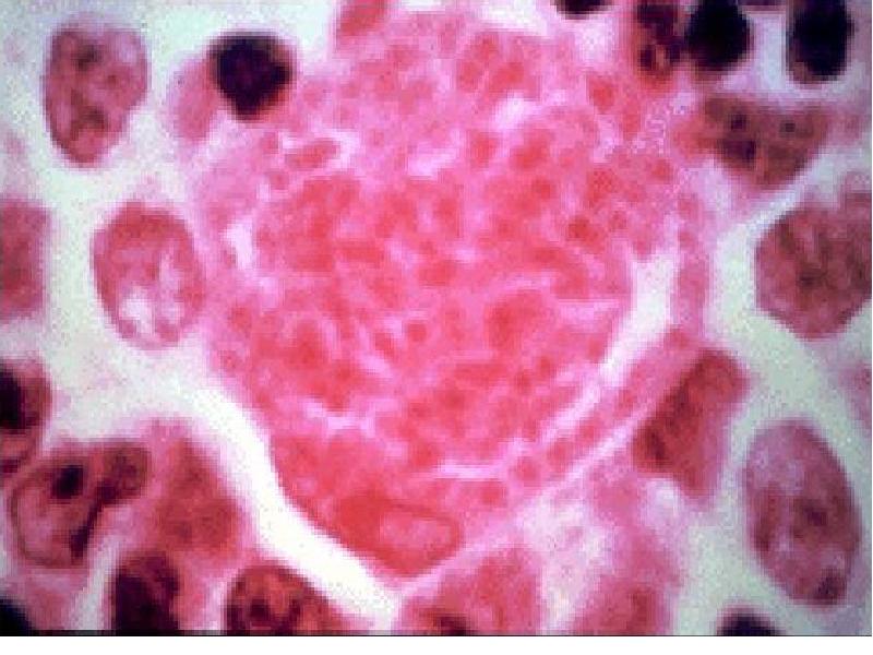
A nuclear cell before destroy ( “false cyst”)
Слайд 13
Описание слайда:
After destroy a cell Т. penetrate into other cell recycling developments. Only at this stage of the parasitemia develops which allows them through a placenta a fetus!!
The cycle of an asexual reproduction is prolonged before creation of immunity, that carries on to termination of a parasitemia and creation tissue cysts in a sceletal muscles, eyes, brain, cardiac muscle. A size cyst up to 100 microns with a dense sheath. Т. in its are as bradyzoites.
Derivation tissue cysts - ending stage biotic cycle Т. in a body of the intermediate host. At this stage the parasitemia is not observed.
At a stage trophozoites Т. are very unstable and fast perish at warming up to 50 d.C., at effect 50 % of alcohol and anyone disinfectants through 5-10 minutes
Слайд 14
Описание слайда:
Слайд 15
Описание слайда:
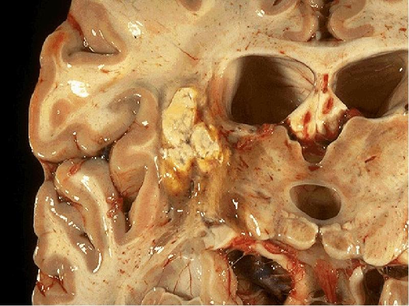
PATHOMORPHOLOGY
The pathological changes are found out in all bodies both at congenital and at the acquired toxoplasmosis.
Congenital Т. CNS - is accompanied by damage retinas and of the choroidinal membranes of the eyes (99 %)
Infectious agents find out as tissue cysts is more often in muscles.
The inflammation around of them misses or is expressed unsignificantly.
Tachyzoites in a blood it are found out only at primary infection.
Слайд 16
Описание слайда:
It is possible to detect granulomas, centers of a necrosis and fibrosis more often in muscles, myocardium, lungs, liver, spleen, lymphatic nodi.
The sites calcification in a brain find only at congenital as semilunars in the field of a striatal body.
The lingering character of an infection results in various allergic manifestations.
The alive Т. can will be saved In cysts all life!
Слайд 17
Описание слайда:
EPIDEMIOLOGY
(zoonosis with peroral and vertical mode of infection)
Infection of the man occurs at:
- contact to the cats and subjects enclosing them
- the use it is not enough heat treated or crude meat
- through a placenta (frequency congenital Т. – 1 : 2,700 labors)
- hemotransfusions and the transplantetions of donor organs
From the man to the man at contact - does not transmit!!!
The proportion infectious of the people coincides with frequency of infection Т. for animals. More often meets in the countries a hot and wet climate
Слайд 18
Описание слайда:
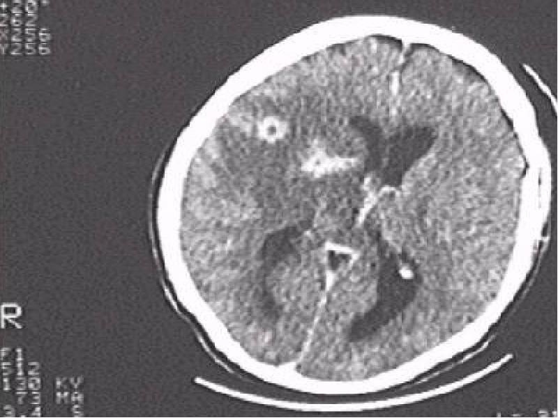
CLINIC (incubation interval about 2 weeks)
Distinguish 2 forms Т. – congenital and acquired
Acquired Т. - acute, chronic, primary or secondary latent)
Acute acquired Т. – it is generalized infection
- acute beginning, fever, intoxication, pain in muscles
the increase of a liver and spleen (rare) is
- polymorphic exanthema by duration 3 - 4 days
- generalized lymphadenopathy
encephalitis with form of parasite abscesses ( CT-scan
or MRI
- pneumonia
- myocarditis
- one-sided chorioretinitis (1 %)
Слайд 19
Описание слайда:
Слайд 20
Описание слайда:
Слайд 21
Описание слайда:
To frequency there is a combination of numbered mani-festations by an extent of several days or weeks spontaneous convalescence. The frequency of lethal outcomes is unknown. The exact diagnosis more often is installed only immunological methods.
Chronic acquired Т. It is long-lived flaccid disease
- subfebrile condition long-lived (months) sometimes with
by periods normal temperature
- chronic intoxication
- damage of many bodies and systems:
- complains: common weakness, lowering of appetite,
dryness in an oral cavity, nausea,
- irritability, disturbance of dream, headache, lowering of
memory,
- pain in heart and palpitation, pain in muscles and joints,
disorder of vision
Слайд 22
Описание слайда:
Objective:
- generalized lymphadenopathy
- increase of a liver (50 %) and spleen
- specific myosites with calcifications
- arthralgias
- tachycardia, hypotension, the phenomena of a myocarditis with focal changes a ECG
GIT of dull pains in epigastriums, abdominal distention,
constipations, weight loss
- NS - emotional lability, lowering of capacity for work, irritability, cancerophobia etc.
Sometimes severe neurosises, diencephalic syndrome,
the symptomatic epilepsy (rare) is and for all vegeto-
vascular disorders
Слайд 23
Описание слайда:
- eyes - chorioretinitis (1 %), uveitis, progressing myopia
- ES - disorders of menstrual cycle, impotence, secondary adrenal unsufficiency, the depression of function of thyroid glands (rare)
- WBC - leukopenia, lymphocytosis, rise eosinocytes at normal values ESR
The most often form of disease is primary latent Т:
- is not present and was not in the past of clinic Т.
Diagnosis only by detection of antibodies against Т. the class Ig G.
Peakings more often only immunological with appearance IgM
The secondary latent Т. - residual phenomena after transferred Т. as calcifications, seams after a chorioretinitis, lowering of vision, scleroses of lymphatic nodi.
Слайд 24
Описание слайда:
CONGENITAL Т. - arises only at primary infection by the pregnant woman.
The gravity of current depends on a duration of gestation in period infection - the less age of a fetus, the heavy the current Т.
Though infection during pregnancy not always results in a congenital toxoplasmosis.
The long-lived overseeing by 176 pregnant woman with primary infection without clinical manifestations has not detected signs Т in 110 children (63 %), 6 - has perished prenatal or during labors and congenital Т. is detected for 30 children, and for 11 children the infection was doubtful.
At severe current Т. the fetus perishes or is born prematurely (31 %). The lethality among newborn thus in 2 times was higher in matching with children is born in time.
Слайд 25
Описание слайда:
Signs of disease is possible to detect for child at once or after many days after labors as:
- fever, lowering of mass of a body
- spotty - papular eruptions on a skin
- generalized lymphadenopathy
- increase of a liver and spleen, appearance of an icterus
- hydrocephalia, microcephalia (50 %), microphthalmia
- psychomotor disability (56 %)
- cramps both common or separate groups of muscles
- chorioretinitis for 99 % (bilateral for 85 %)
- the detection of intracranial calcifications - is more often
in a striatal body of a brain as of semilunar lines.
The acute infection Т. usually results in lethal outcome within the first days or weeks after labors
Слайд 26
Описание слайда:
But it can pass in the chronic or secondary - latent forms Т., having left after itself:
- microphthalmia, hydrocephalus
- chorioretinitis, paralyses of eye’s muscles
- psychosomatic or motive disability, cramps.
These children are needed in medical observation and
constant control, as the true damage to health newborn is
possible to estimate more often only after some weeks or
months etc.
Слайд 27
Описание слайда:
DIFFERENTIAL DIAGNOSTICS will be carried out by :
- Catscratch disease
- Herpes simplex
- Histoplasmosis
- Listeriosis
- Cytomegalovirus
- HIV-infection, Pneumocytosis
- Infectious mononucleosis,
- Lymphoreticulosis, lymphogranulomatosis,sarcoidosis, Lymphoma
- Sepsis,
- Thyrotoxicosis, chronic tonsillitis etc.
- Rheumatic disease, myocardites,
- Syphilis,
- Mesadenitis
- Hemolytic illness (at congenital Т.),
- Tuberculosis
Слайд 28
Описание слайда:
LABORATORY DIAGNOSIS.
1.Microscopy smears CSF ( at congenital Т.) – xanthochromatic, rise eosinocytes and protein
2. Microscopy smears of a blood, bioptates of lymphatic nodi, tonsils, placenta, membranes of a fetus, amniotic fluid (search cysts and Т.)
3. Biological test with the same material with the subsequent search cysts in sections of tissues animal
4. PCR - CSF, amniotic waters, tissues of the perished fetus, placenta etc.
5. Immunological methods – ELISA, CFt, IHAt
The IC test with toxoplasmin now will not be used
Слайд 29
Описание слайда:
TREATMENT
The antiparasitic therapy
Will not be carried out:
- at chronic Т. outside of peaking,
- at is primary also secondary - latent Т.
Will be carried out:
at acute Т.,
chronic toxoplasmosis with clinical manifestations,
at a damage of a brain and eyes. using:
- Pyrimethamine 50 - 100 mg 1 po- first day-main drug
then on 25 –50mg 1 po - 29 days
- Sulfadiazine 1-1.5 gm po q6h
Слайд 30
Описание слайда:
for strengthen operation of pyrimethaminum:
- clindamycin 300 mg po/IV q6h
- claritromycin 1 gm po bid
- azithromycin 1.2-1.5 gm po qd
Spiramycine 1gm po q8h (for the pregnant
women) - is less effective of pyrimethaminum
- Leucovorin 10mg 1 po or 1gm of fresh beer yeast po
(for elimination of a side effect of medicines: leukopenia,
thrombocytopenia, anemia)
Слайд 31
Описание слайда:
For the pregnant woman at immunological peaking without clinical manifestations - drugs "of the «HEEL" corporation of Germany now will be used
They have no contraindications and do not render terato-genic of operation under the particular scheme with usage: Echinacea-composite, Coenzyme - composite, Nozode of a toxoplasmosis, Lymphomyosote, Galiume or Psorinoheel etc.
The drugs should be selected by a method of medicamental
testing. The efficiency of treatment makes 93 % ( disappearence of Ig M from of blood of pregnant woman )
Besides the symptomatic and pathogenetic therapy will be carried out.
Слайд 32
Описание слайда:
PROPHYLAXIS
The special attention accesses on group of hazard among the pregnant woman (absence in plasma of antibodies against Т.) They should interrupt contacts to the cats, and also use meat a past valuable heat treatment, carefully wash hands before eating etc.)
Group of hazard in a population owes revealed among the girls since 16 years and before pregnancy - valuable inspection on ТОRCH- of an infection here should be carried out and during pregnancy to carry out definition of antibodies only Ig M to the detected earlier infections from group TORCH (HSH, CMV, ТОХО, rubella)
Woman, which up to pregnancy have antibodies against Т. – give birth to healthy children.
Other measures because of a wide circulation of a toxoplasmosis - have small efficiency!!
Слайд 33
Описание слайда:
Скачать презентацию на тему TOXOPLASMOSIS ( T.) Identification. Zoonotic disease caused by an о bligate можно ниже: