Chapter 42 презентация
Содержание
- 2. Overview: Trading Places Every organism must exchange materials with its environment.
- 3. For most cells making up multicellular organisms, direct exchange with the
- 5. Circulatory systems link exchange surfaces with cells throughout the body In
- 6. Gastrovascular Cavities Simple animals, such as cnidarians, have a body wall
- 8. Open and Closed Circulatory Systems More complex animals have either open
- 9. In insects, other arthropods, and most molluscs, blood bathes the organs
- 10. In a closed circulatory system, the blood is confined to vessels
- 12. Organization of Vertebrate Closed Circulatory Systems Humans and other vertebrates have
- 13. Arteries branch into arterioles and carry blood to capillaries. Arteries
- 14. Vertebrate hearts contain two or more chambers. Vertebrate hearts contain two
- 15. Single Circulation Bony fishes, rays, and sharks have single circulation with
- 17. Double Circulation Amphibian, reptiles, and mammals have double circulation. Oxygen-poor and
- 19. In reptiles and mammals, oxygen-poor blood flows through the pulmonary circuit
- 20. Adaptations of Double Circulatory Systems Amphibians: Frogs / amphibians have a
- 21. Reptiles (Except Birds) Turtles, snakes, and lizards have a three-chambered heart:
- 22. Mammals Mammals and birds have a four-chambered heart with two atria
- 23. Coordinated cycles of heart contraction drive double circulation in mammals Blood
- 24. Blood returns to the heart through the superior vena cava (deoxygenated
- 26. The Mammalian Heart: A Closer Look A closer look at the
- 28. The heart contracts and relaxes in a rhythmic cycle called the
- 30. The heart rate, also called the pulse, is the number of
- 31. Four valves prevent backflow of blood in the heart: Four valves
- 32. Maintaining the Heart’s Rhythmic Beat Some cardiac muscle cells are self-excitable
- 34. Patterns of blood pressure and flow reflect the structure and arrangement
- 36. Capillaries have thin walls, the endothelium plus its basement membrane, to
- 37. Blood Flow Velocity Physical laws governing movement of fluids through pipes
- 39. Blood Pressure Blood pressure is the hydrostatic pressure that blood exerts
- 40. Changes in Blood Pressure During the Cardiac Cycle Systolic pressure is
- 41. Regulation of Blood Pressure Blood pressure is determined by cardiac output
- 42. Vasoconstriction and vasodilation help maintain adequate blood flow as the body’s
- 45. Fainting is caused by inadequate blood flow to the head. Fainting
- 47. Capillary Function Capillaries in major organs are usually filled to capacity.
- 49. The critical exchange of substances between the blood and interstitial fluid
- 51. Fluid Return by the Lymphatic System The lymphatic system - returns
- 52. Lymph nodes are organs that produce phagocytic white blood cells and
- 53. Blood Composition and Function Blood consists of several kinds of blood
- 55. Plasma Blood plasma is about 90% water. Among its solutes are
- 56. Cellular Elements Suspended in blood plasma are two types of cells:
- 57. Red blood cells, or erythrocytes, are by far the most numerous
- 58. Leukocytes - Defense There are five major types of white blood
- 59. Platelets - Blood Clotting Platelets are fragments of cells and function
- 61. Stem Cells and the Replacement of Cellular Elements The cellular elements
- 63. Cardiovascular Disease = Disorders of the Heart and the Blood Vessels
- 65. Treatment and Diagnosis of Cardiovascular Disease Cholesterol is a major contributor
- 66. Gas exchange occurs across specialized respiratory surfaces Gas exchange supplies oxygen
- 67. Respiratory Media Animals can use air or water as a source
- 68. Respiratory Surfaces Animals require large, moist respiratory surfaces for exchange of
- 70. Ventilation moves the respiratory medium over the respiratory surface. Ventilation moves
- 72. Tracheal Systems in Insects The tracheal system of insects consists of
- 74. Lungs = Infoldings of the body surface The circulatory system
- 75. Mammalian Respiratory Systems: A Closer Look A system of branching ducts
- 77. Breathing Ventilates the Lungs by Inhalation and Exhalation of Air
- 79. How a Bird Breathes Birds have eight or nine air sacs
- 81. Control of Breathing in Humans In humans, the main breathing control
- 82. Sensors in the aorta and carotid arteries monitor O2 and CO2
- 84. Adaptations for gas exchange include pigments that bind and transport gases
- 86. Respiratory Pigments Respiratory pigments = proteins that transport oxygen, greatly increase
- 87. Hemoglobin A single hemoglobin molecule can carry four molecules of O2
- 90. Carbon Dioxide Transport Hemoglobin also helps transport CO2 and assists in
- 92. Elite Animal Athletes Migratory and diving mammals have evolutionary adaptations that
- 94. You should now be able to: Compare and contrast open and
- 95. Define cardiac cycle and explain the role of the sinoatrial node.
- 96. Describe the role played by the lymphatic system in relation to
- 97. For humans, describe the exchange of gases in the lungs and
- 98. Скачать презентацию


Turtles, snakes, and lizards have a three-chambered heart:")


Слайды и текст этой презентации
Слайд 1
Описание слайда:
Chapter 42
Circulation and Gas Exchange
Слайд 2
Описание слайда:
Overview: Trading Places
Every organism must exchange materials with its environment.
Exchanges ultimately occur at the cellular level.
In unicellular organisms, these exchanges occur directly with the environment.
Слайд 3
Описание слайда:
For most cells making up multicellular organisms, direct exchange with the environment is not possible.
For most cells making up multicellular organisms, direct exchange with the environment is not possible.
Gills are an example of a specialized exchange system in animals.
Internal transport and gas exchange are functionally related in most animals.
Слайд 4
Описание слайда:
Слайд 5
Описание слайда:
Circulatory systems link exchange surfaces with cells throughout the body
In small and/or thin animals, cells can exchange materials directly with the surrounding medium.
In most animals, transport systems connect the organs of exchange with the body cells.
Most complex animals have internal transport systems that circulate fluid.
Слайд 6
Описание слайда:
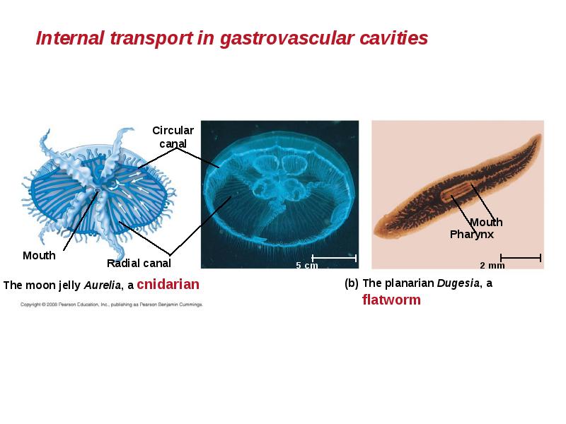
Gastrovascular Cavities
Simple animals, such as cnidarians, have a body wall that is only two cells thick and that encloses a gastrovascular cavity.
This cavity functions in both digestion and distribution of substances throughout the body.
Some cnidarians, such as jellies, have elaborate gastrovascular cavities.
Flatworms have a gastrovascular cavity and a large surface area to volume ratio.
Слайд 7
Описание слайда:
Слайд 8
Описание слайда:
Open and Closed Circulatory Systems
More complex animals have either open or closed circulatory systems.
Both systems have three basic components:
A circulatory fluid = blood or hemolymph.
A set of tubes = blood vessels.
A muscular pump = the heart.
Слайд 9
Описание слайда:
In insects, other arthropods, and most molluscs, blood bathes the organs directly in an open circulatory system.
In insects, other arthropods, and most molluscs, blood bathes the organs directly in an open circulatory system.
In an open circulatory system, there is no distinction between blood and interstitial fluid, and this general body fluid is more correctly called hemolymph.
Слайд 10
Описание слайда:
In a closed circulatory system, the blood is confined to vessels and is distinct from the interstitial fluid.
In a closed circulatory system, the blood is confined to vessels and is distinct from the interstitial fluid.
Closed systems are more efficient at transporting circulatory fluids to tissues and cells.
Слайд 11
Описание слайда:
Слайд 12
Описание слайда:
Organization of Vertebrate Closed Circulatory Systems
Humans and other vertebrates have a closed circulatory system, often called the cardiovascular system.
The three main types of blood vessels are:
arteries - away from the heart.
veins - toward the heart.
capillaries - exchange with body cells.
Слайд 13
Описание слайда:
Arteries branch into arterioles and carry blood to capillaries.
Arteries branch into arterioles and carry blood to capillaries.
Networks of capillaries called capillary beds are the sites of chemical exchange between the blood and interstitial fluid.
Venules converge into veins and return blood from capillaries to the heart.
Слайд 14
Описание слайда:
Vertebrate hearts contain two or more chambers.
Vertebrate hearts contain two or more chambers.
Blood enters through an atrium and is pumped out through a ventricle.
Atria - receive blood
Ventricles - pump blood
Слайд 15
Описание слайда:
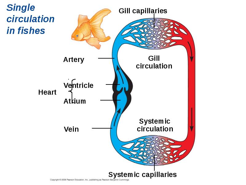
Single Circulation
Bony fishes, rays, and sharks have single circulation with a two-chambered heart.
In single circulation, blood leaving the heart passes through two capillary beds before returning.
Слайд 16
Описание слайда:
Слайд 17
Описание слайда:
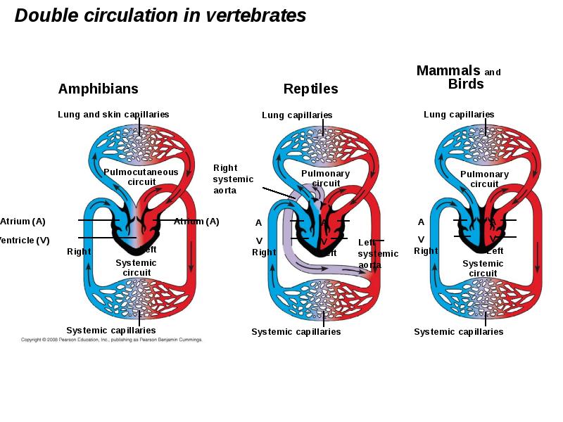
Double Circulation
Amphibian, reptiles, and mammals have double circulation.
Oxygen-poor and oxygen-rich blood are pumped separately from the right and left sides of the heart.
Слайд 18
Описание слайда:
Слайд 19
Описание слайда:
In reptiles and mammals, oxygen-poor blood flows through the pulmonary circuit to pick up oxygen through the lungs.
In reptiles and mammals, oxygen-poor blood flows through the pulmonary circuit to pick up oxygen through the lungs.
In amphibians, oxygen-poor blood flows through a pulmocutaneous circuit to pick up oxygen through the lungs and skin.
Oxygen-rich blood delivers oxygen through the systemic circuit.
Double circulation maintains higher blood pressure in the organs than does single circulation.
Слайд 20
Описание слайда:
Adaptations of Double Circulatory Systems
Amphibians:
Frogs / amphibians have a three-chambered heart: 2 atria and 1 ventricle.
The ventricle pumps blood into a forked artery that splits the ventricle’s output into the pulmocutaneous circuit and the systemic circuit.
Underwater, blood flow to the lungs is nearly shut off.
Слайд 21
Описание слайда:
Reptiles (Except Birds)
Turtles, snakes, and lizards have a three-chambered heart: two atria and one ventricle.
In alligators, caimans, and other crocodilians a septum - partially or fully divides the ventricle.
Reptiles have double circulation, with a pulmonary circuit - lungs and a systemic circuit.
Слайд 22
Описание слайда:
Mammals
Mammals and birds have a four-chambered heart with two atria and two ventricles.
The left side of the heart pumps and receives only oxygen-rich blood, while the right side receives and pumps only oxygen-poor blood.
Mammals and birds are endotherms and require more O2 than ectotherms.
Слайд 23
Описание слайда:
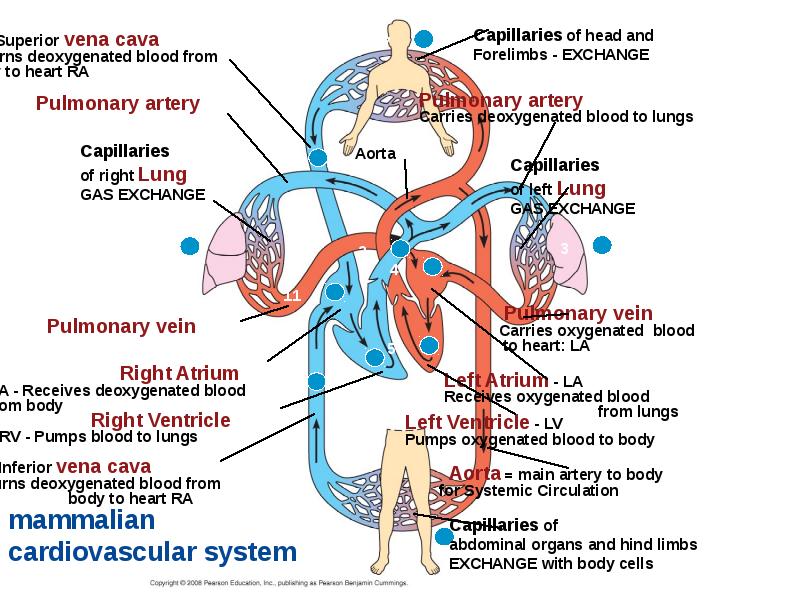
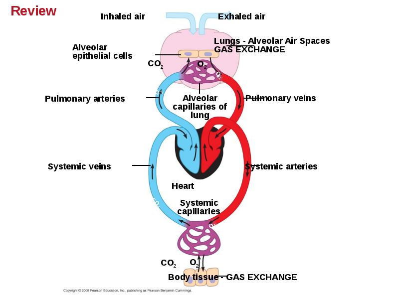
Coordinated cycles of heart contraction drive double circulation in mammals
Blood begins its flow with the right ventricle pumping blood to the lungs.
In the lungs, the blood loads O2 and unloads CO2
Oxygen-rich blood from the lungs enters the heart at the left atrium and is pumped through the aorta to the body tissues by the left ventricle.
The aorta provides blood to the heart through the coronary arteries.
Слайд 24
Описание слайда:
Blood returns to the heart through the superior vena cava (deoxygenated blood from head, neck, and forelimbs) and inferior vena cava (deoxygenated blood from trunk and hind limbs).
Blood returns to the heart through the superior vena cava (deoxygenated blood from head, neck, and forelimbs) and inferior vena cava (deoxygenated blood from trunk and hind limbs).
The superior vena cava and inferior vena cava flow into the Right Atrium - RA.
Слайд 25
Описание слайда:
Слайд 26
Описание слайда:
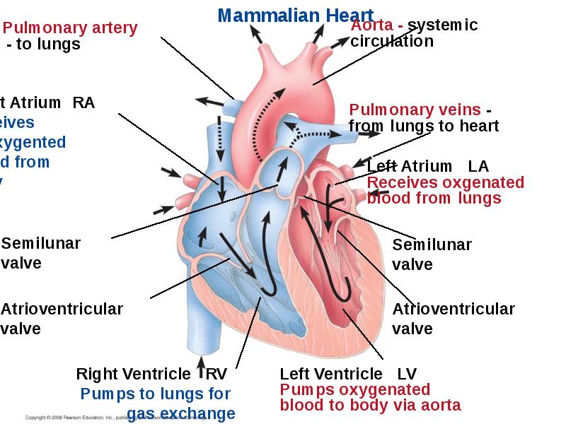
The Mammalian Heart: A Closer Look
A closer look at the mammalian heart provides a better understanding of double circulation.
RIGHT side = deoxygenated blood from body pumped to lungs.
LUNGS = gas exchange.
LEFT side = oxygenated blood from lungs pumped to body.
Слайд 27
Описание слайда:
Слайд 28
Описание слайда:
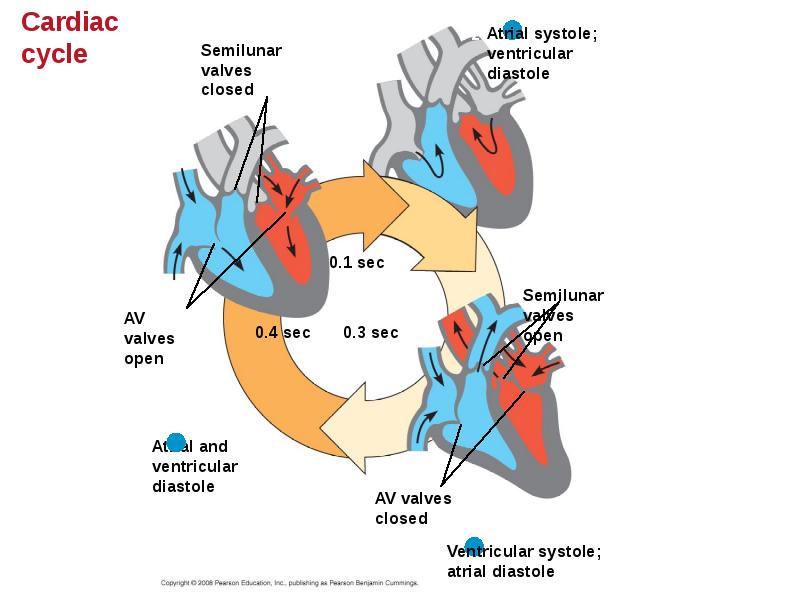
The heart contracts and relaxes in a rhythmic cycle called the cardiac cycle.
The heart contracts and relaxes in a rhythmic cycle called the cardiac cycle.
The contraction, or pumping, phase is called systole.
The relaxation, or filling, phase is called diastole.
Blood Pressure = systolic / diastolic
Слайд 29
Описание слайда:
Слайд 30
Описание слайда:
The heart rate, also called the pulse, is the number of beats per minute.
The heart rate, also called the pulse, is the number of beats per minute.
The stroke volume is the amount of blood pumped in a single contraction.
The cardiac output is the volume of blood pumped into the systemic circulation per minute and depends on both the heart rate and stroke volume.
Слайд 31
Описание слайда:
Four valves prevent backflow of blood in the heart:
Four valves prevent backflow of blood in the heart:
The atrioventricular (AV) valves separate each atrium and ventricle.
The semilunar valves control blood flow to the aorta and the pulmonary artery.
The “lub-dup” sound of a heart beat is caused by the recoil of blood against the AV valves (lub) then against the semilunar (dup) valves.
Backflow of blood through a defective valve causes a heart murmur.
Слайд 32
Описание слайда:
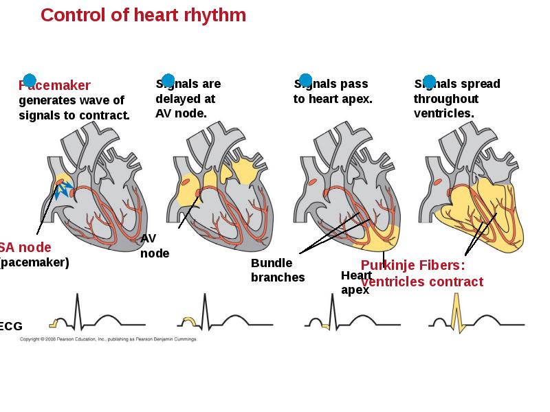
Maintaining the Heart’s Rhythmic Beat
Some cardiac muscle cells are self-excitable = they contract without any signal from the nervous system.
The sinoatrial (SA) node, or pacemaker, sets the rate and timing at which cardiac muscle cells contract.
Impulses from the SA node travel to the atrioventricular (AV) node. At the AV node, the impulses are delayed and then travel to the Purkinje fibers that make the ventricles contract.
Impulses that travel during the cardiac cycle can be recorded as an electrocardiogram (ECG or EKG). The pacemaker is influenced by nerves, hormones, body temperature, and exercise.
Слайд 33
Описание слайда:
Слайд 34
Описание слайда:
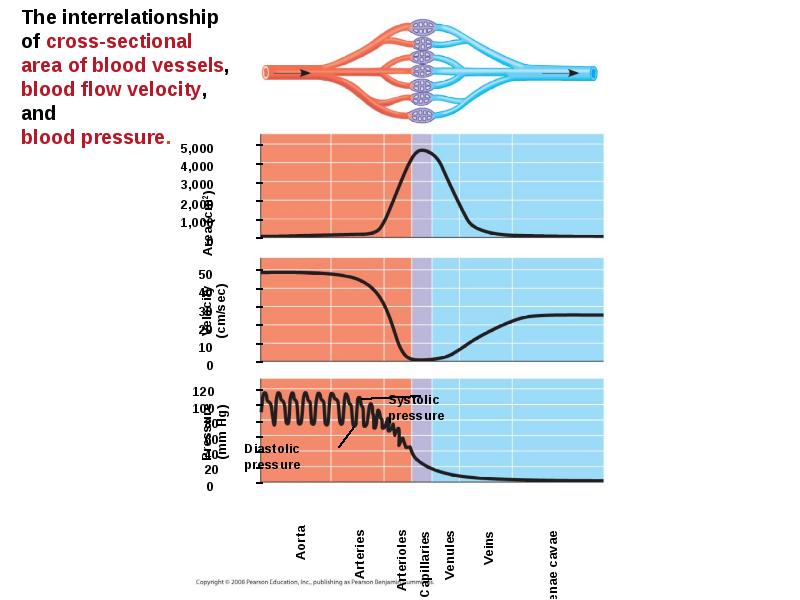
Patterns of blood pressure and flow reflect the structure and arrangement of blood vessels
The physical principles that govern movement of water in plumbing systems also influence the functioning of animal circulatory systems.
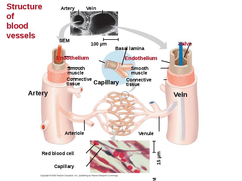
The epithelial layer that lines blood vessels is called the endothelium.
Слайд 35
Описание слайда:
Слайд 36
Описание слайда:
Capillaries have thin walls, the endothelium plus its basement membrane, to facilitate the exchange of materials.
Capillaries have thin walls, the endothelium plus its basement membrane, to facilitate the exchange of materials.
Arteries and veins have an endothelium, smooth muscle, and connective tissue.
Arteries have thicker walls than veins to accommodate the high pressure of blood pumped from the heart.
In the thinner-walled veins, blood flows back to the heart mainly as a result of muscle action.
Слайд 37
Описание слайда:
Blood Flow Velocity
Physical laws governing movement of fluids through pipes affect blood flow and blood pressure.
Velocity of blood flow is slowest in the capillary beds, as a result of the high resistance and large total cross-sectional area.
Blood flow in capillaries is necessarily slow for exchange of materials.
Слайд 38
Описание слайда:
Слайд 39
Описание слайда:
Blood Pressure
Blood pressure is the hydrostatic pressure that blood exerts against the wall of a vessel.
In rigid vessels blood pressure is maintained; less rigid vessels deform and blood pressure is lost.
Слайд 40
Описание слайда:
Changes in Blood Pressure During the Cardiac Cycle
Systolic pressure is the pressure in the arteries during ventricle contraction /systole; it is the highest pressure in the arteries.
Diastolic pressure is the pressure in the arteries during relaxation /diastole; it is lower than systolic pressure.
A pulse is the rhythmic bulging of artery walls with each heartbeat.
Слайд 41
Описание слайда:
Regulation of Blood Pressure
Blood pressure is determined by cardiac output and peripheral resistance due to constriction of arterioles.
Vasoconstriction is the contraction of smooth muscle in arteriole walls; it increases blood pressure.
Vasodilation is the relaxation of smooth muscles in the arterioles; it causes blood pressure to fall.
Слайд 42
Описание слайда:
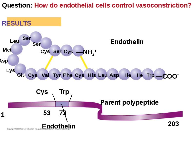
Vasoconstriction and vasodilation help maintain adequate blood flow as the body’s demands change.
Vasoconstriction and vasodilation help maintain adequate blood flow as the body’s demands change.
The peptide endothelin is an important inducer of vasoconstriction.
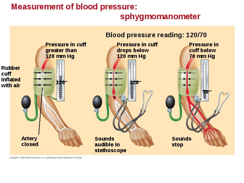
Blood pressure is generally measured for an artery in the arm at the same height as the heart.
Blood pressure for a healthy 20 year old at rest is 120 mm Hg at systole / 70 mm Hg at diastole.
Слайд 43
Описание слайда:
Слайд 44
Описание слайда:
Слайд 45
Описание слайда:
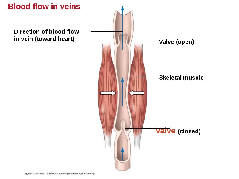
Fainting is caused by inadequate blood flow to the head.
Fainting is caused by inadequate blood flow to the head.
Animals with longer necks require a higher systolic pressure to pump blood a greater distance against gravity.
Blood is moved through veins by smooth muscle contraction, skeletal muscle contraction, and expansion of the vena cava with inhalation.
One-way valves in veins / heart prevent backflow of blood.
Слайд 46
Описание слайда:
Слайд 47
Описание слайда:
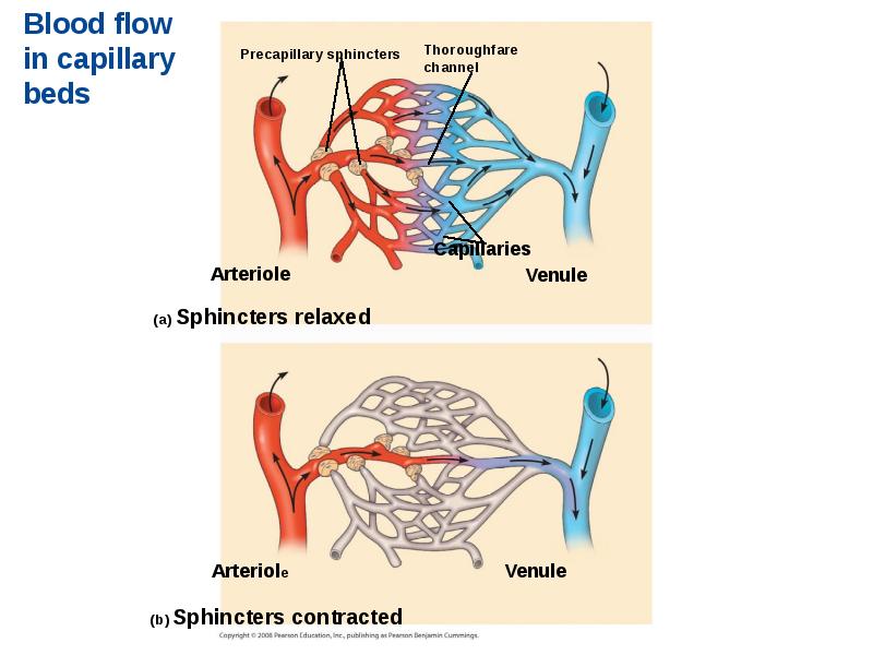
Capillary Function
Capillaries in major organs are usually filled to capacity. Blood supply varies in many other sites.
Two mechanisms regulate distribution of blood in capillary beds:
Contraction of the smooth muscle layer in the wall of an arteriole constricts the vessel.
Precapillary sphincters control flow of blood between arterioles and venules.
Слайд 48
Описание слайда:
Слайд 49
Описание слайда:
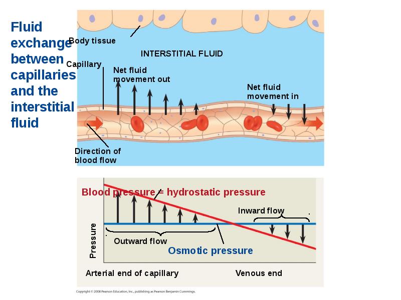
The critical exchange of substances between the blood and interstitial fluid takes place across the thin endothelial walls of the capillaries.
The critical exchange of substances between the blood and interstitial fluid takes place across the thin endothelial walls of the capillaries.
The difference between blood pressure and osmotic pressure drives fluids out of capillaries at the arteriole end and into capillaries at the venule end.
Слайд 50
Описание слайда:
Слайд 51
Описание слайда:
Fluid Return by the Lymphatic System
The lymphatic system - returns fluid that leaks out in the capillary beds … restoring filtered fluid to blood maintains homeostasis.
This system aids in body defense.
Fluid, called lymph, reenters the circulation directly at the venous end of the capillary bed and indirectly through the lymphatic system.
The lymphatic system drains into neck veins.
Слайд 52
Описание слайда:
Lymph nodes are organs that produce phagocytic white blood cells and filter lymph - an important role in the body’s defense.
Lymph nodes are organs that produce phagocytic white blood cells and filter lymph - an important role in the body’s defense.
Edema is swelling caused by disruptions in the flow of lymph.
Слайд 53
Описание слайда:
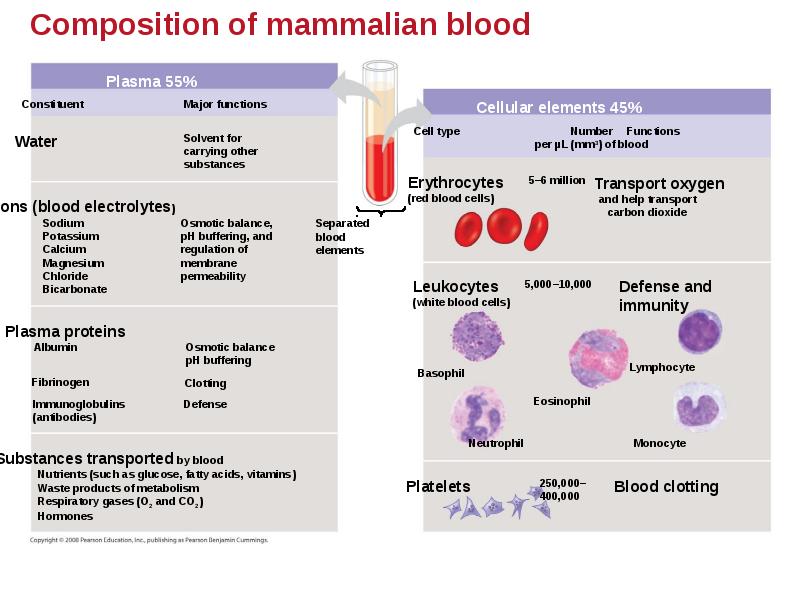
Blood Composition and Function
Blood consists of several kinds of blood cells suspended in a liquid matrix called plasma.
The cellular elements: red blood cells, white blood cells, and platelets occupy about 45% of the volume of blood.
Слайд 54
Описание слайда:
Слайд 55
Описание слайда:
Plasma
Blood plasma is about 90% water.
Among its solutes are inorganic salts in the form of dissolved ions, sometimes called electrolytes.
Another important class of solutes is the plasma proteins, which influence blood pH, osmotic pressure, and viscosity. Various plasma proteins function in lipid transport, immunity, and blood clotting.
Plasma transports nutrients, gases, and cell waste.
Слайд 56
Описание слайда:
Cellular Elements
Suspended in blood plasma are two types of cells:
Red blood cells rbc = erythrocytes, transport oxygen.
White blood cells wbc = leukocytes, function in defense.
Platelets are fragments of cells that are involved in blood clotting.
Слайд 57
Описание слайда:
Red blood cells, or erythrocytes, are by far the most numerous blood cells.
They transport oxygen throughout the body.
They contain hemoglobin, the iron-containing protein that transports oxygen.
Слайд 58
Описание слайда:
Leukocytes - Defense
There are five major types of white blood cells, or leukocytes: monocytes, neutrophils, basophils, eosinophils, and lymphocytes.
They function in defense by phagocytizing bacteria and debris or by producing antibodies.
They are found both in and outside of the circulatory system.
Слайд 59
Описание слайда:
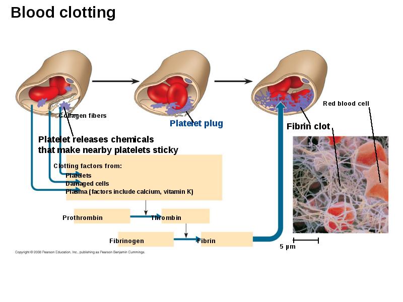
Platelets - Blood Clotting
Platelets are fragments of cells and function in blood clotting.
When the endothelium of a blood vessel is damaged, the clotting mechanism begins.
A cascade of complex reactions converts fibrinogen to fibrin, forming a clot.
A blood clot formed within a blood vessel is called a thrombus and can block blood flow.
Слайд 60
Описание слайда:
Слайд 61
Описание слайда:
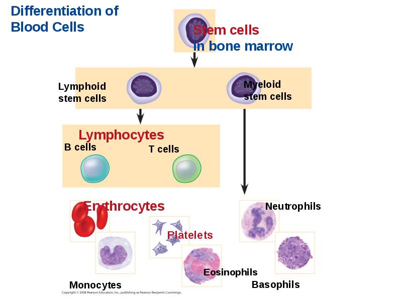
Stem Cells and the Replacement of Cellular Elements
The cellular elements of blood wear out and are replaced constantly throughout a person’s life.
Erythrocytes, leukocytes, and platelets all develop from a common source of stem cells in the red marrow of bones.
The hormone erythropoietin (EPO) stimulates erythrocyte production when oxygen delivery is low.
Слайд 62
Описание слайда:
Слайд 63
Описание слайда:
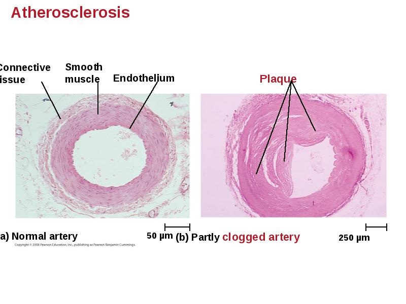
Cardiovascular Disease = Disorders of the Heart and the Blood Vessels
One type of cardiovascular disease, atherosclerosis, is caused by the buildup of plaque deposits within arteries.
A heart attack is the death of cardiac muscle tissue resulting from blockage of one or more coronary arteries.
A stroke is the death of nervous tissue in the brain, usually resulting from rupture or blockage of arteries in the brain /head.
Слайд 64
Описание слайда:
Слайд 65
Описание слайда:
Treatment and Diagnosis of Cardiovascular Disease
Cholesterol is a major contributor to atherosclerosis.
Low-density lipoproteins (LDLs) = “bad cholesterol,” are associated with plaque formation.
High-density lipoproteins (HDLs) = “good cholesterol,” reduce the deposition of cholesterol.
Hypertension = high blood pressure, promotes atherosclerosis and increases the risk of heart attack and stroke.
Hypertension can be reduced by dietary changes, exercise, and/or medication.
Слайд 66
Описание слайда:
Gas exchange occurs across specialized respiratory surfaces
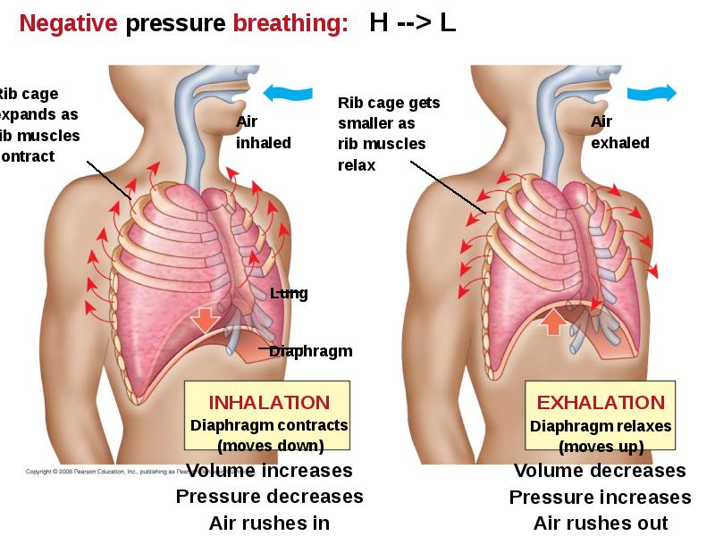
Gas exchange supplies oxygen for cellular respiration and disposes of carbon dioxide. Gases diffuse down pressure gradients in the lungs and other organs as a result of differences in partial pressure.
Partial pressure is the pressure exerted by a particular gas in a mixture of gases. A gas diffuses from a region of higher partial pressure to a region of lower partial pressure: H --> L
In the lungs and tissues, O2 and CO2 diffuse from where their partial pressures are higher to where they are lower.
Слайд 67
Описание слайда:
Respiratory Media
Animals can use air or water as a source of O2, or respiratory medium.
In a given volume, there is less O2 available in water than in air.
Obtaining O2 from water requires greater efficiency than air breathing.
Слайд 68
Описание слайда:
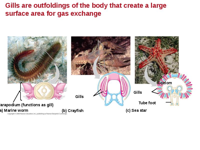
Respiratory Surfaces
Animals require large, moist respiratory surfaces for exchange of gases between their cells and the respiratory medium, either air or water.
Gas exchange across respiratory surfaces takes place by diffusion.
Respiratory surfaces vary by animal and can include the outer surface, skin, gills, tracheae, and lungs.
Слайд 69
Описание слайда:
Слайд 70
Описание слайда:
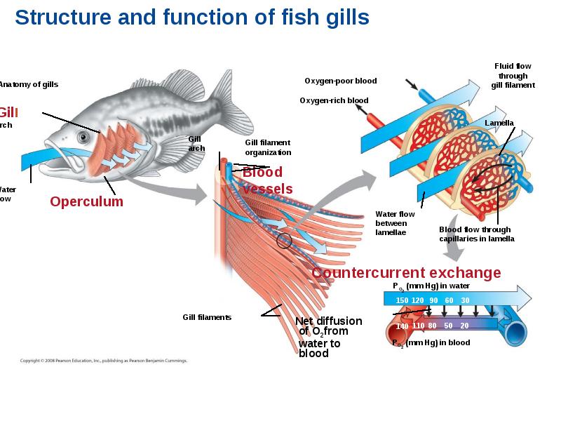
Ventilation moves the respiratory medium over the respiratory surface.
Ventilation moves the respiratory medium over the respiratory surface.
Aquatic animals move through water or move water over their gills for ventilation.
Fish gills use a countercurrent exchange system, where blood flows in the opposite direction to water passing over the gills; blood is always less saturated with O2 than the water it meets… maximizes diffusion.
Слайд 71
Описание слайда:
Слайд 72
Описание слайда:
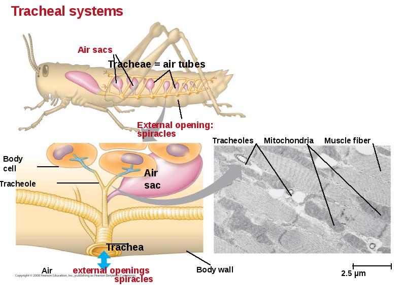
Tracheal Systems in Insects
The tracheal system of insects consists of tiny branching tubes that penetrate the body.
The tracheal tubes supply O2 directly to body cells.
The respiratory and circulatory systems are separate.
Larger insects must ventilate their tracheal system to meet O2 demands.
Слайд 73
Описание слайда:
Слайд 74
Описание слайда:
Lungs = Infoldings of the body surface
The circulatory system (open or closed) transports gases between the lungs and the rest of the body.
The size and complexity of lungs correlate with an animal’s metabolic rate.
Слайд 75
Описание слайда:
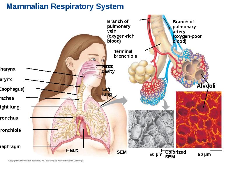
Mammalian Respiratory Systems: A Closer Look
A system of branching ducts / air tubes conveys air to the lungs.
Air inhaled through the nostrils --> pharynx --> larynx --> trachea --> bronchi --> bronchioles --> alveoli = site of gas exchange.
Exhaled air passes over the vocal cords to create sounds.
Alveoli are wrapped by capillaries for GAS EXCHANGE.
Слайд 76
Описание слайда:
Слайд 77
Описание слайда:
Breathing Ventilates the Lungs by Inhalation and
Exhalation of Air
Amphibians, such as a frog, ventilates its lungs by positive pressure breathing, which forces air down the trachea.
Mammals ventilate by negative pressure breathing, which pulls air into the lungs by varying volume / air pressure. Lung volume increases as the rib muscles and diaphragm contract.
The tidal volume is the volume of air inhaled with each breath. The maximum tidal volume is the vital capacity. After exhalation, residual volume of air remains in the lungs.
Слайд 78
Описание слайда:
Слайд 79
Описание слайда:
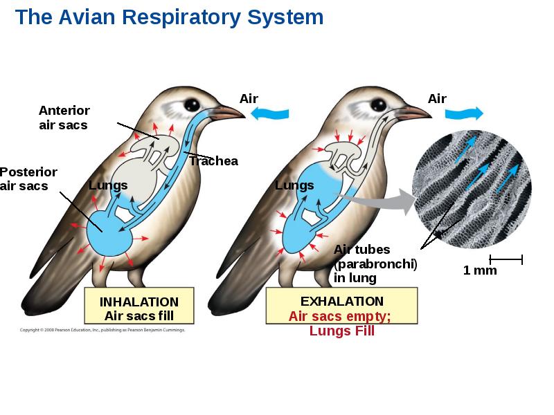
How a Bird Breathes
Birds have eight or nine air sacs that function as bellows that keep air flowing through the lungs.
Air passes through the lungs in one direction only.
Every exhalation completely renews the air in the lungs.
Слайд 80
Описание слайда:
Слайд 81
Описание слайда:
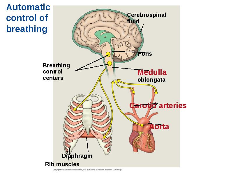
Control of Breathing in Humans
In humans, the main breathing control centers are in two regions of the brain, the medulla oblongata and the pons.
The medulla regulates the rate and depth of breathing in response to pH changes - CO2 levels in the cerebrospinal fluid.
The medulla adjusts breathing rate and depth to match metabolic demands.
The pons regulates the tempo.
Слайд 82
Описание слайда:
Sensors in the aorta and carotid arteries monitor O2 and CO2 concentrations in the blood.
Sensors in the aorta and carotid arteries monitor O2 and CO2 concentrations in the blood.
These sensors exert secondary control over breathing.
Слайд 83
Описание слайда:
Слайд 84
Описание слайда:
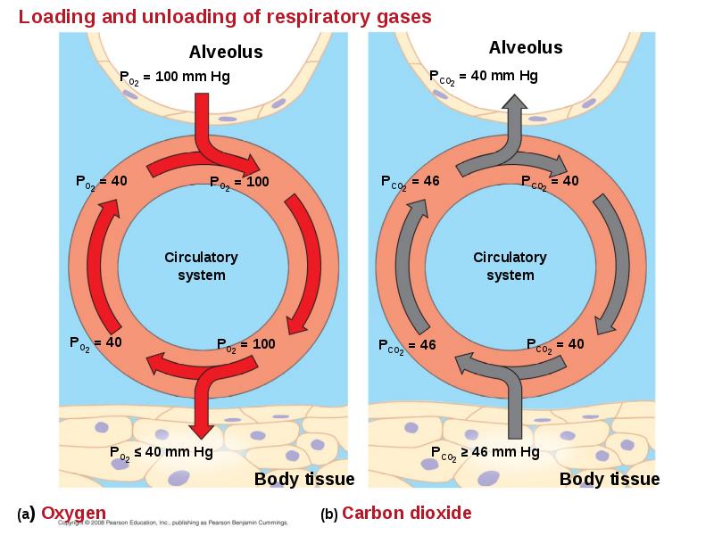
Adaptations for gas exchange include pigments that bind and transport gases
The metabolic demands of many organisms require that the blood transport large quantities of O2 and CO2
Blood arriving in the lungs has a low partial pressure of O2 and a high partial pressure of CO2 relative to air in the alveoli.
In the alveoli, O2 diffuses into the blood and CO2 diffuses into the air.
In tissue capillaries, partial pressure gradients favor diffusion of O2 into the interstitial fluids and CO2 into the blood.
Слайд 85
Описание слайда:
Слайд 86
Описание слайда:
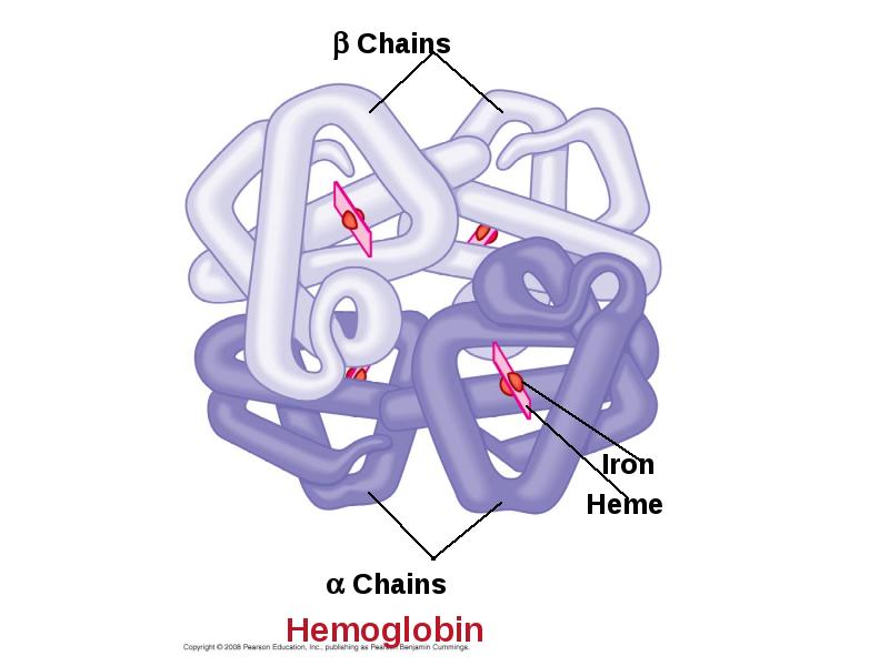
Respiratory Pigments
Respiratory pigments = proteins that transport oxygen, greatly increase the amount of oxygen that blood can carry.
Arthropods and many molluscs have hemocyanin with copper as the oxygen-binding component.
Most vertebrates and some invertebrates use hemoglobin with iron = oxygen-binding component contained within erythrocytes.
Слайд 87
Описание слайда:
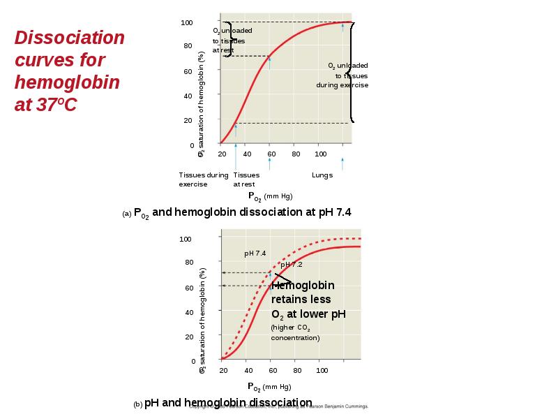
Hemoglobin
A single hemoglobin molecule can carry four molecules of O2
The hemoglobin dissociation curve shows that a small change in the partial pressure of oxygen can result in a large change in delivery of O2
CO2 produced during cellular respiration lowers blood pH and decreases the affinity of hemoglobin for O2
This is called the Bohr shift.
Слайд 88
Описание слайда:
Слайд 89
Описание слайда:
Слайд 90
Описание слайда:
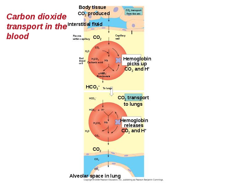
Carbon Dioxide Transport
Hemoglobin also helps transport CO2 and assists in buffering.
CO2 from respiring cells diffuses into the blood and is transported either in blood plasma, bound to hemoglobin, or as bicarbonate ions = HCO3–.
Слайд 91
Описание слайда:
Слайд 92
Описание слайда:
Elite Animal Athletes
Migratory and diving mammals have evolutionary adaptations that allow them to perform extraordinary feats.
The extreme O2 consumption of the antelope-like pronghorn underlies its ability to run at high speed over long distances.
Deep-diving air breathers stockpile O2 and deplete it slowly.
Weddell seals have a high blood to body volume ratio and can store oxygen in their muscles in myoglobin proteins.
Слайд 93
Описание слайда:
Слайд 94
Описание слайда:
You should now be able to:
Compare and contrast open and closed circulatory systems.
Compare and contrast the circulatory systems of fish, amphibians, reptiles, and mammals or birds.
Distinguish between pulmonary and systemic circuits and explain the function of each.
Trace the path of a red blood cell through the human heart, pulmonary circuit, and systemic circuit.
Слайд 95
Описание слайда:
Define cardiac cycle and explain the role of the sinoatrial node.
Define cardiac cycle and explain the role of the sinoatrial node.
Relate the structures of capillaries, arteries, and veins to their function.
Define blood pressure and cardiac output and describe two factors that influence each.
Explain how osmotic pressure and hydrostatic pressure regulate the exchange of fluid and solutes across the capillary walls.
Слайд 96
Описание слайда:
Describe the role played by the lymphatic system in relation to the circulatory system.
Describe the role played by the lymphatic system in relation to the circulatory system.
Describe the function of erythrocytes, leukocytes, platelets, fibrin.
Distinguish between a heart attack and stroke.
Discuss the advantages and disadvantages of water and of air as respiratory media.
Слайд 97
Описание слайда:
For humans, describe the exchange of gases in the lungs and in tissues.
For humans, describe the exchange of gases in the lungs and in tissues.
Draw and explain the hemoglobin-oxygen dissociation curve.
Скачать презентацию на тему Chapter 42 можно ниже: