Introduction in topographic anatomy and operative surgery презентация
Содержание
- 2. Topographical anatomy is a science about the dimensional structure of healthy
- 3. The operative surgery is a science about surgical operations, methods of
- 4. M.I. Pyrogov ( 1810-1881)
- 5. Classification of operations Emergency Urgent Planned Bloodless Bloody Radical Palliative
- 6. Operative approach means to make the wound for the exposure of
- 7. Operative method – the main part of the operation, performing the
- 8. General surgical instruments Scalpels
- 9. Positions of scalpels, forceps а —scalpels; 1 — position of bow;
- 10. The scissors
- 11. The surgical saw
- 12. Forceps
- 13. Retractors
- 14. Instruments for the arrest bleeding
- 15. Needles
- 16. Suture material Absorbable Plain catgut Chromic catgut Polyglycolic synthetics
- 17. Type of sutures Interrupted Continuous
- 18. Regions of the Head and Neck
- 20. Layer Structure of Fronto-parieto-occipital Region Skin; subcutaneous tissues; gala aponeurotica; loose
- 22. Arterial and nerve supply of the Scalp The supratrochlear and the
- 24. The venous drainage of the Scalp The supratrochlear and supraorbital veins
- 26. Temporal region and parotid regions
- 27. Layer Structure of Temporal Region Skin; subcutaneous tissues; temporal aponeurosis:
- 28. The four arteries anastomose on the inferior surface of the brain
- 29. Internal base the skull,dura mater,venous sinuses and cranial nerves
- 31. Decompression trepanation
- 34. Potential places of intracranial hematoma
- 37. Скачать презентацию


")


Слайды и текст этой презентации
Слайд 1
Описание слайда:
Introduction in topographic anatomy and operative surgery
Associate-professor Slabyy O.B.
Слайд 2
Описание слайда:
Topographical anatomy is a science about the dimensional structure of healthy human body organs, tissues and parts of the body
Слайд 3
Описание слайда:
The operative surgery is a science about surgical operations, methods of surgical operations, the essence of which comes to mechanical action upon the organs and tissues with diagnostic, medical or reconstructive purpose.
Слайд 4
Описание слайда:
M.I. Pyrogov ( 1810-1881)
Слайд 5
Описание слайда:
Classification of operations
Emergency
Urgent
Planned
Bloodless
Bloody
Radical
Palliative
Слайд 6
Описание слайда:
Operative approach means to make the wound for the exposure of the organ to be operated on
Слайд 7
Описание слайда:
Operative method – the main part of the operation, performing the action contained in the name of the operation
Слайд 8
Описание слайда:
General surgical instruments
Scalpels
Слайд 9
Описание слайда:
Positions of scalpels, forceps
а —scalpels; 1 — position of bow; 2 — position of table knife; 3 —writing pen; 4 — amputating knife; б — forceps
Слайд 10
Описание слайда:
The scissors
Слайд 11
Описание слайда:
The surgical saw
Слайд 12
Описание слайда:
Forceps
Слайд 13
Описание слайда:
Retractors
Слайд 14
Описание слайда:
Instruments for the arrest bleeding
Слайд 15
Описание слайда:
Needles
Слайд 16
Описание слайда:
Suture material
Absorbable
Plain catgut
Chromic catgut
Polyglycolic synthetics
Слайд 17
Описание слайда:
Type of sutures
Interrupted
Continuous
Слайд 18
Описание слайда:
Regions of the Head and Neck
Слайд 19
Описание слайда:
Слайд 20
Описание слайда:
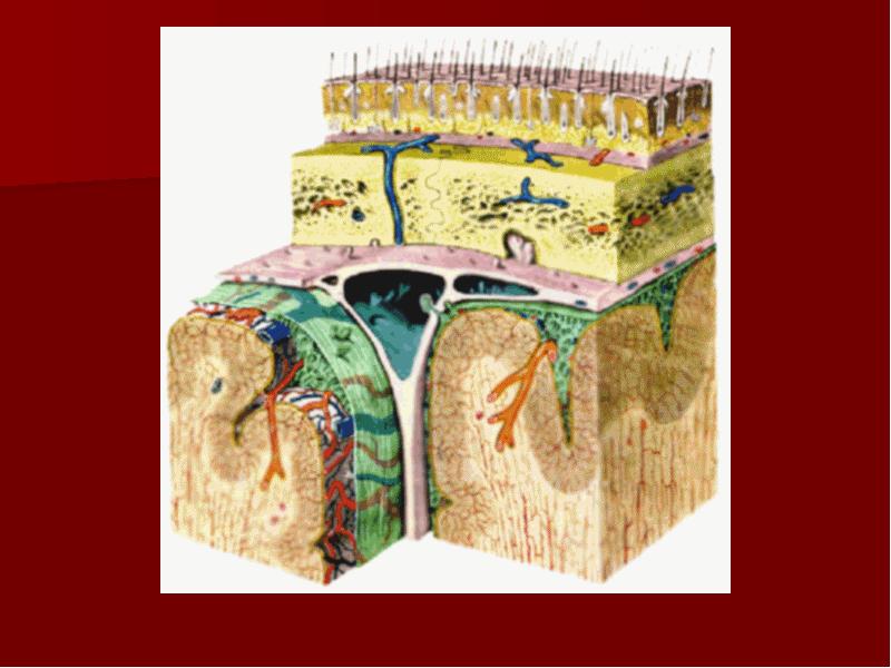
Layer Structure of Fronto-parieto-occipital Region
Skin;
subcutaneous tissues;
gala aponeurotica;
loose areolar tissue;
periosteum (pericranium);
loose areolar tissue;
bone (internal, external lamina and diploe).
Слайд 21
Описание слайда:
Слайд 22
Описание слайда:
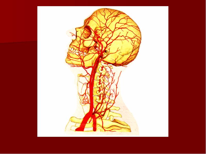
Arterial and nerve supply of the Scalp
The supratrochlear and the supraorbital arteries in company with supratrochlear and the supraorbital nerves.
The superficial temporal artery,zygomaticotemporal and auriculotemporal nerve.
The posterior auricular artery and lesser occipital nerve (cervical plexus C2)
The occiptal artery and greater occipital nerve (posterior ramus of the second cervical nerve).
Слайд 23
Описание слайда:
Слайд 24
Описание слайда:
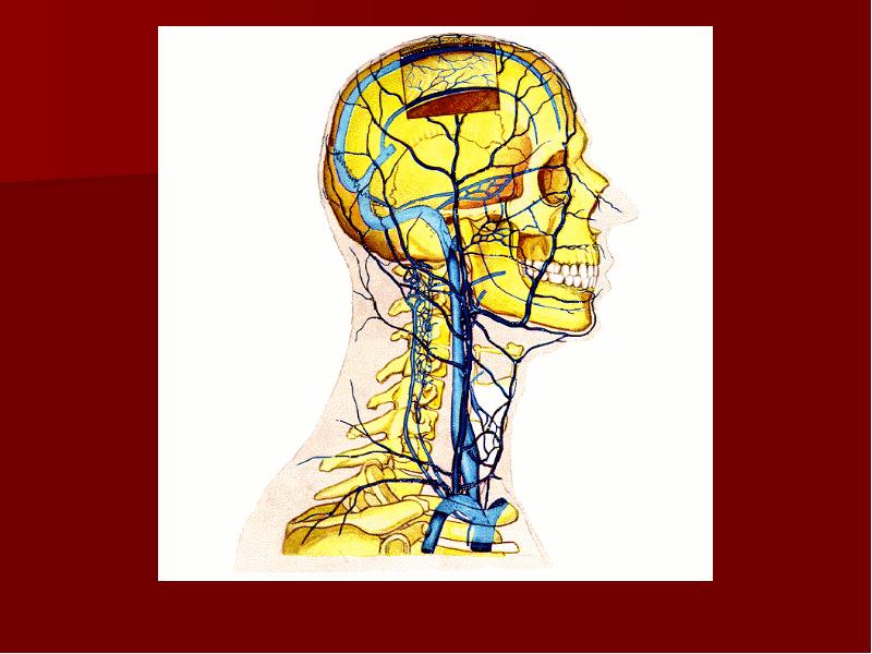
The venous drainage of the Scalp
The supratrochlear and supraorbital veins (to from the facial vein).
The superficial temporal vein (to from the retromandibular vein).
The postrior auricular vein (to from the external jugular vein).
The occipital vein (into the suboccipital venous plexus, in turn into the vertebral veins, occasionally forward into the internal jugular vein.
The veins of the Scalp freely anastomose with another and are connected to the diploic veins and the intracranial venous sinuses by the valveless emissary veins.
Слайд 25
Описание слайда:
Слайд 26
Описание слайда:
Temporal region and parotid regions
Слайд 27
Описание слайда:
Layer Structure of Temporal Region
Skin;
subcutaneous tissues;
temporal aponeurosis:
- external lamina;
- loose areolar tissue;
- internal lamina;
4. subaponeurotical fat;
5. temporal muscle;
6. submuscular loose areolar tissue;
7. pericranium;
8. temporal bone.
Слайд 28
Описание слайда:
The four arteries anastomose on the inferior surface of the brain and form the circulus arteriosus
Слайд 29
Описание слайда:
Internal base the skull,dura mater,venous sinuses and cranial nerves
Слайд 30
Описание слайда:
Слайд 31
Описание слайда:
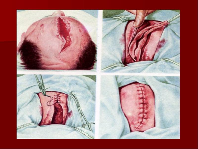
Decompression trepanation
Слайд 32
Описание слайда:
Слайд 33
Описание слайда:
Слайд 34
Описание слайда:
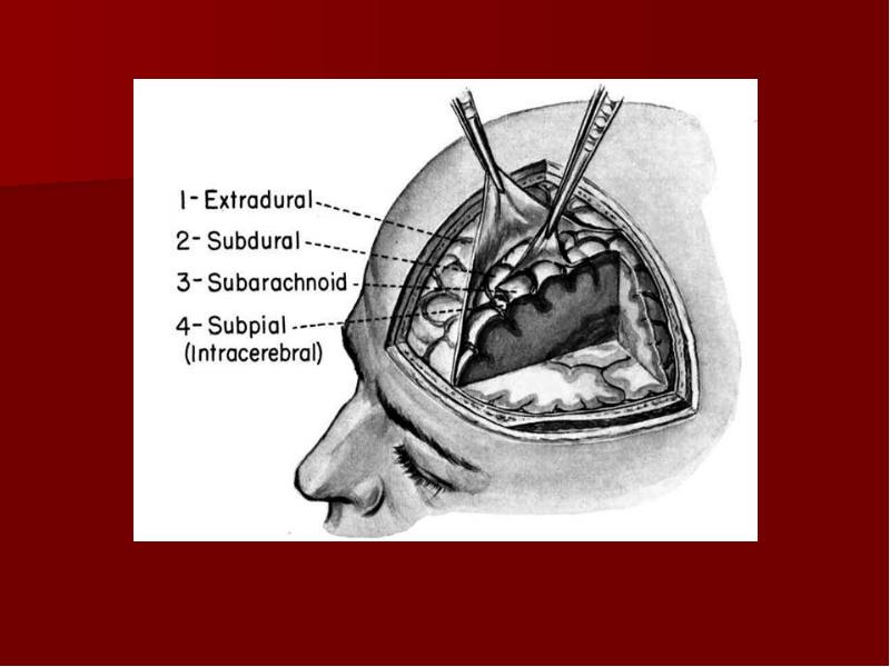
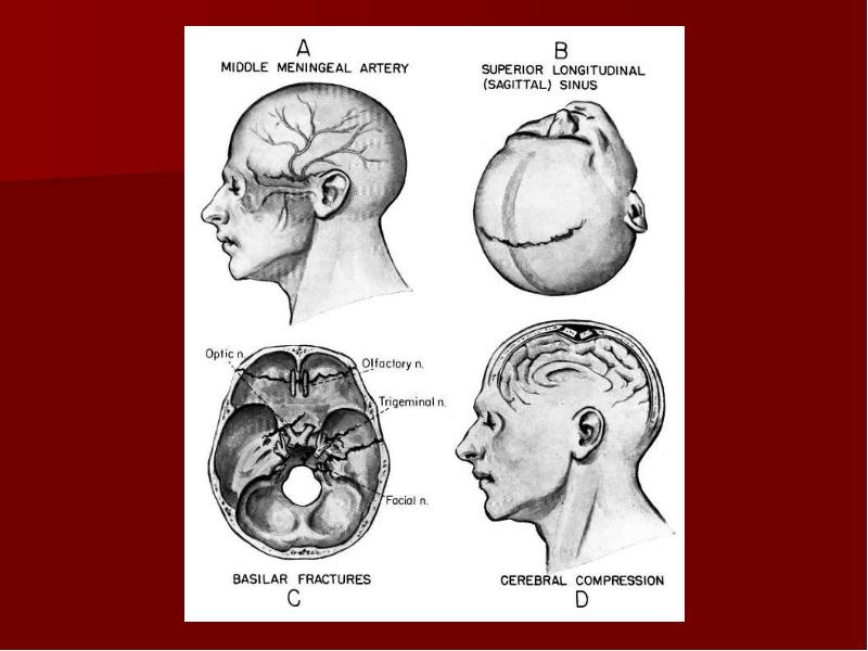
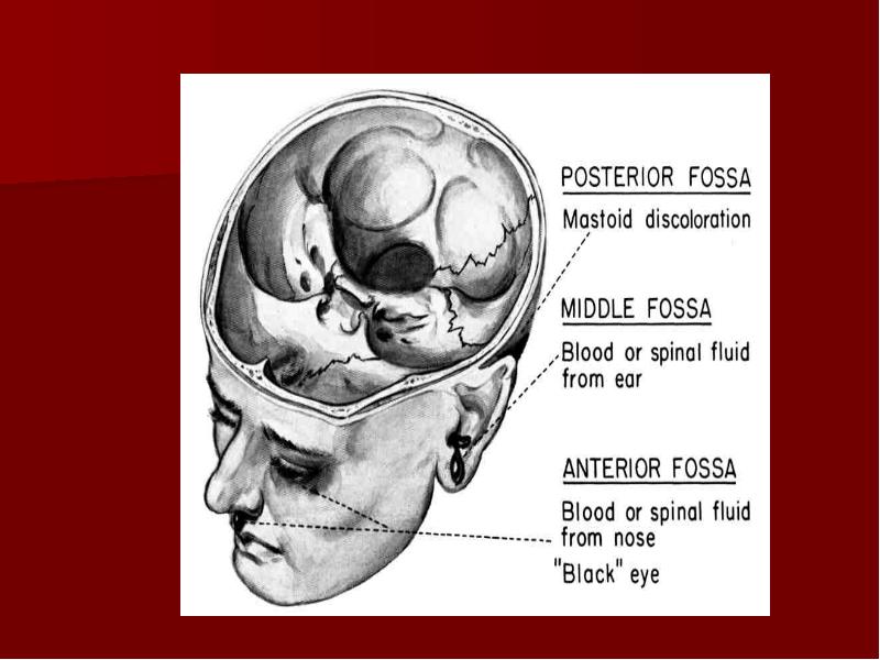
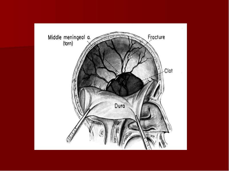
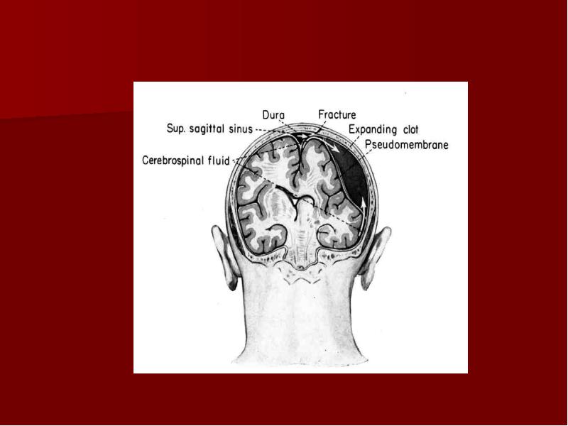
Potential places of intracranial hematoma
Слайд 35
Описание слайда:
Слайд 36
Описание слайда:
Скачать презентацию на тему Introduction in topographic anatomy and operative surgery можно ниже: