Lung Examination: Abnormal презентация
Содержание
- 5. Illustrative Pathological problems Consolidation Atelectasis Pleural effusion Pneumothorax Mass Diffuse lung
- 12. Steps General Examination Mediastinal position Chest expansion Lung resonance Breath sounds
- 13. General Examination Respiratory rate Pattern of breathing Cyanosis Clubbing Weight Cough
- 14. Respiratory Rate Bradypnea: rate less than 8 per minute Tachypnea:
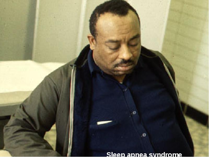
- 15. Pattern of Breathing Kussmals Sleep apnea Cheyne strokes Pursed lip breathing
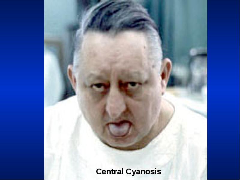
- 17. Central Cyanosis Results from pulmonary dysfunction, the mucous membrane of conjunctiva
- 21. Clubbing In clubbing, there is widening of the AP and lateral
- 22. Significance: Clubbing Observed In: Intrathoracic malignancy: Primary or secondary (lung, pleural,
- 24. Weight Emaciation cachectic Malignancy Tuberculosis
- 26. Weight Obese: Sleep apnea syndrome
- 28. Cough Productive Dry Whooping Bovine
- 30. Hospital Setting Isolation room Oxygen set up
- 31. Effort of Ventilation Person appears uncomfortable. Breathing seems voluntary. Accessory muscles
- 32. Resting Size and Shape of Thorax Barrel chest Kyphosis Scoliosis Pectus
- 33. Barrel Chest
- 34. Tracheal Position: Mediastinum Any deviation of the mediastinum is abnormal Lateral
- 36. Chest Expansion Asymmetrical chest expansion is abnormal The abnormal side expands
- 37. Percussion: Decreased or Increased Resonance is Abnormal Dullness Decreased resonance is
- 38. Breath Sounds: Diminished or Absent Intensity of breath sounds, in general,
- 39. Bronchial Bronchial breathing anywhere other than over the trachea, right clavicle
- 40. Bronchial breathing
- 41. Rhonchi Rhonchi are long continuous adventitious sounds, generated by obstruction to
- 42. Rhonchi
- 43. Rhonchi Localized rhonchi suggests obstruction of any etiology e.g., tumor, foreign
- 44. Pleural Rub Normal parietal and visceral pleura glide smoothly during respiration.
- 45. Pleural rub
- 46. Stridor Loud audible inspiratory rhonchi is called a stridor. Inspiratory rhonchi
- 48. Crackles Interrupted adventitious sounds are called crackles. Make a notation about
- 49. Crackles When the crackles are heard at the end of inspiration
- 50. Voice Transmission (tactile fremitus, vocal resonance) Asymmetrical voice transmission points to
- 51. Voice Transmission (tactile fremitus, vocal resonance) Decreased: A quantitative decrease in
- 53. Скачать презентацию


Asymmetrical voice transmission points to")

Decreased: A quantitative decrease in")
Слайды и текст этой презентации
Слайд 1
Описание слайда:
Lung Examination: Abnormal
Arcot J. Chandrasekhar, M.D.
Слайд 2
Описание слайда:
Слайд 3
Описание слайда:
Слайд 4
Описание слайда:
Слайд 5
Описание слайда:
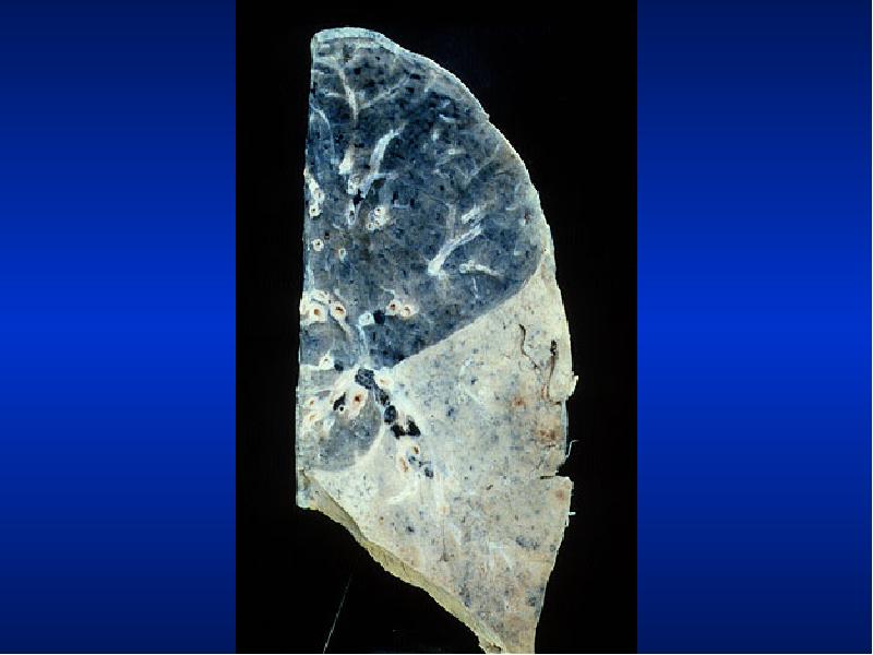
Illustrative Pathological problems
Consolidation
Atelectasis
Pleural effusion
Pneumothorax
Mass
Diffuse lung disease
Слайд 6
Описание слайда:
Слайд 7
Описание слайда:
Слайд 8
Описание слайда:
Слайд 9
Описание слайда:
Слайд 10
Описание слайда:
Слайд 11
Описание слайда:
Слайд 12
Описание слайда:
Steps
General Examination
Mediastinal position
Chest expansion
Lung resonance
Breath sounds
Adventitious sounds
Voice transmission
Слайд 13
Описание слайда:
General Examination
Respiratory rate
Pattern of breathing
Cyanosis
Clubbing
Weight
Cough
Hospital setting
Effort of ventilation
Shape of thorax
Слайд 14
Описание слайда:
Respiratory Rate
Bradypnea: rate less than 8 per minute
Tachypnea: rate greater than 25 per minute
Слайд 15
Описание слайда:
Pattern of Breathing
Kussmals
Sleep apnea
Cheyne strokes
Pursed lip breathing
Orthopnoea: Short of breath in supine position, gets some relief by sitting or standing up.
Слайд 16
Описание слайда:
Слайд 17
Описание слайда:
Central Cyanosis
Results from pulmonary dysfunction, the mucous membrane of conjunctiva and tongue are bluish.
If there was chronic hypoxemia and secondary erythrocytosis, you can detect the conjunctival and scleral vessels to be full, tortuous and bluish.
Слайд 18
Описание слайда:
Слайд 19
Описание слайда:
Слайд 20
Описание слайда:
Слайд 21
Описание слайда:
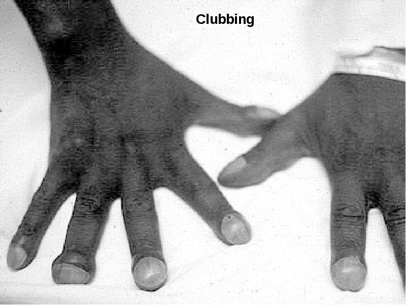
Clubbing
In clubbing, there is widening of the AP and lateral diameter of terminal portion of fingers and toes giving the appearance of clubbing.
The angle between the nail and skin is greater than 180.
The periungual skin is stretched and shiny.
There is fluctuation of the nail bed.
One can feel the posterior edge of the nail.
Слайд 22
Описание слайда:
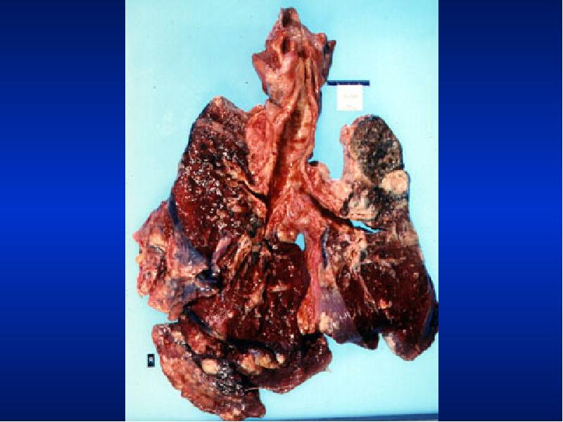
Significance: Clubbing Observed In:
Intrathoracic malignancy: Primary or secondary (lung, pleural, mediastinal)
Suppurative lung disease: (lung abscess, bronchiectasis, empyema)
Diffuse interstitial fibrosis: Alveolar capillary block syndrome
In association with other systemic disorders
Слайд 23
Описание слайда:
Слайд 24
Описание слайда:
Weight
Emaciation cachectic
Malignancy
Tuberculosis
Слайд 25
Описание слайда:
Слайд 26
Описание слайда:
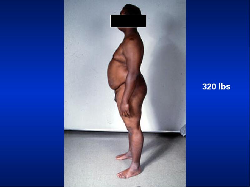
Weight
Obese: Sleep apnea syndrome
Слайд 27
Описание слайда:
Слайд 28
Описание слайда:
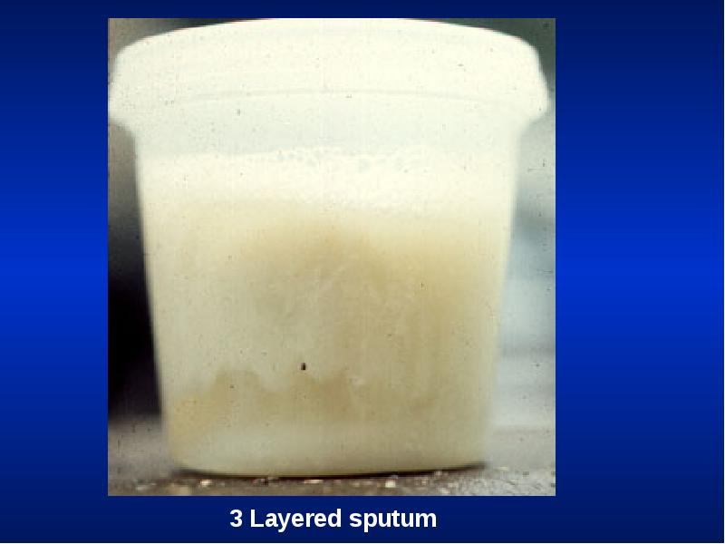
Cough
Productive
Dry
Whooping
Bovine
Слайд 29
Описание слайда:
Слайд 30
Описание слайда:
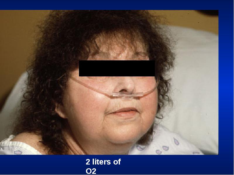
Hospital Setting
Isolation room
Oxygen set up
Слайд 31
Описание слайда:
Effort of Ventilation
Person appears uncomfortable. Breathing seems voluntary.
Accessory muscles are in use, expiratory muscles are active and expiration is not passive any more.
The degree of negative pleural pressure is high.
The respiratory rate is increased.
Слайд 32
Описание слайда:
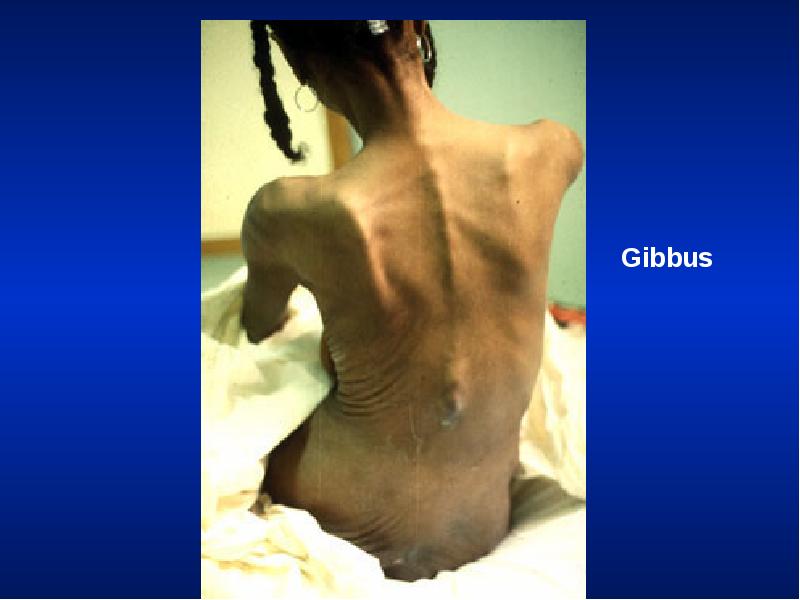
Resting Size and Shape of Thorax
Barrel chest
Kyphosis
Scoliosis
Pectus excavatum
Gibbus
Слайд 33
Описание слайда:
Barrel Chest
Слайд 34
Описание слайда:
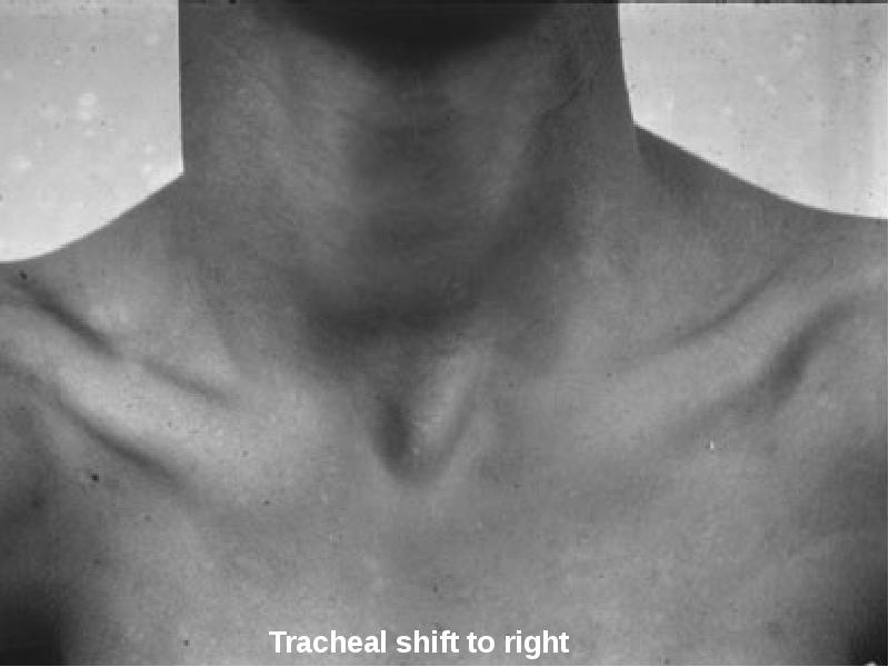
Tracheal Position: Mediastinum
Any deviation of the mediastinum is abnormal
Lateral shift: The mediastinum can be either pulled or pushed away from the lesion
Pull: Loss of lung volume (Atelectasis, fibrosis, agenesis, surgical resection, pleural fibrosis)
Push: Space occupying lesions (pleural effusion, pneumothorax, large mass lesions)
Mediastinal masses and thyroid tumors
Слайд 35
Описание слайда:
Слайд 36
Описание слайда:
Chest Expansion
Asymmetrical chest expansion is abnormal
The abnormal side expands less and lags behind the normal side
Any form of unilateral lung or pleural disease can cause asymmetry of chest expansion
Global expansion decrease
Слайд 37
Описание слайда:
Percussion: Decreased or Increased Resonance is Abnormal
Dullness
Decreased resonance is noted with pleural effusion and all other lung diseases
The dullness is flat and the finger is painful to percussion with pleural effusion
Hyper resonance: Increased resonance can be noted either due to lung distention as seen in asthma, emphysema, bullous disease or due to Pneumothorax
Traube's space
Слайд 38
Описание слайда:
Breath Sounds: Diminished or Absent
Intensity of breath sounds, in general, is a good index of ventilation of the underlying lung.
Breath sounds are markedly decreased in emphysema.
Symmetry: If there is asymmetry in intensity, the side where there is decreased intensity is abnormal.
Any form of pleural or pulmonary disease can give rise to decreased intensity.
Harsh or increased: If the intensity increases there is more ventilation and vice versa.
Слайд 39
Описание слайда:
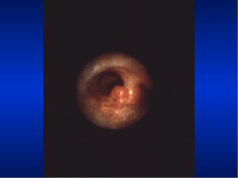
Bronchial
Bronchial breathing anywhere other than over the trachea, right clavicle or right inter-scapular space is abnormal.
In consolidation, the bronchial breathing is low pitched and sticky and is termed tubular type of bronchial breathing.
In cavitary disease, it is high pitched and hollow and is called cavernous breathing. You can simulate this sound by blowing over an empty coke bottle.
Слайд 40
Описание слайда:
Bronchial breathing
Слайд 41
Описание слайда:
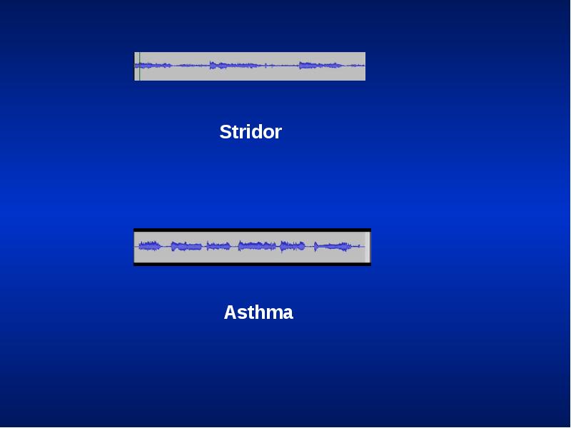
Rhonchi
Rhonchi are long continuous adventitious sounds, generated by obstruction to airways.
When detected, note whether it is generalized or localized, during inspiration or expiration, and the pitch.
Diffused rhonchi would suggest a disease with generalized airway obstruction like asthma or COPD.
Слайд 42
Описание слайда:
Rhonchi
Слайд 43
Описание слайда:
Rhonchi
Localized rhonchi suggests obstruction of any etiology e.g., tumor, foreign body or mucous.
Mucous secretions will disappear with coughing, so would the rhonchus.
Expiratory rhonchi implies obstruction to intrathoracic airways.
Asthmatics can also have inspiratory rhonchi while it is uncommon in COPD.
Слайд 44
Описание слайда:
Pleural Rub
Normal parietal and visceral pleura glide smoothly during respiration.
If the pleura is roughened due to any reason, a scratching, grating sound, related to respiration is heard.
You can hear the sound by compressing harder with the stethoscope and making the patient take deep breaths.
It is localized and can be palpable.
Слайд 45
Описание слайда:
Pleural rub
Слайд 46
Описание слайда:
Stridor
Loud audible inspiratory rhonchi is called a stridor.
Inspiratory rhonchi in general, implies large airway obstruction.
Слайд 47
Описание слайда:
Слайд 48
Описание слайда:
Crackles
Interrupted adventitious sounds are called crackles.
Make a notation about timing, intensity, effect with respiration, position, coughing and character.
Timing and Intensity Crackles heard only at the end of inspiration are called fine crackles.
When the surfactant is depleted, the alveoli collapse. Air enters the alveoli at the end of inspiration.
This sound is generated as the alveoli pop open from it's collapsed state.
Слайд 49
Описание слайда:
Crackles
When the crackles are heard at the end of inspiration and the beginning of expiration the fluid or secretions are probably in respiratory bronchioles: medium crackles.
If the crackles are heard throughout it implies the secretions are in bronchi: coarse crackles.
Слайд 50
Описание слайда:
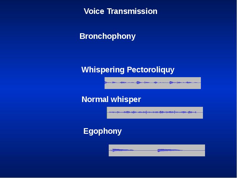
Voice Transmission (tactile fremitus, vocal resonance)
Asymmetrical voice transmission points to disease on one side.
Increased:
Any situation where bronchial breathing is heard the sounds become loud, sharp and distinct: Bronchophony.
In extreme situations, the whispered words come clearly and distinctly: Whispering pectoriloquy.
Слайд 51
Описание слайда:
Voice Transmission (tactile fremitus, vocal resonance)
Decreased: A quantitative decrease in voice transmission could be due to any other form of lung or pleural disease.
Qualitative alteration:
A qualitative alteration of voice transmission is noted over consolidation and along the upper margin of pleural effusion: Egophony
The sound is like a nasal twang or goat bleating.
Слайд 52
Описание слайда:
Скачать презентацию на тему Lung Examination: Abnormal можно ниже: