TAKAYASU’S ARTERITIS презентация
Содержание
- 2. EPIDEMIOLOGY More case reports from Japan ,India, South-east Asia, Mexico No
- 3. Age Age Mc-2nd & 3rd decade May range from
- 4. Genetics Japan - HLA-B52 and B39 Mexican and Colombian patients
- 5. Histopathology Idiopathic c/c infla arteritis of elastic arteries resulting in occlusive
- 7. Gross Gross
- 8. Wall thickening, Fibrosis, Stenosis, & Thrombus formation →end organ ischaemia More
- 9. Associated pathology-TB (LN)-55% Associated pathology-TB (LN)-55%
- 10. Clinical features Early pre pulseless/gen manif
- 12. Coronary involvement in TA Occurs in 10~30% Often fatal Classified into
- 13. Occular involvement-Amaurosis fugax, pain behind eye,
- 16. Ishikawa clinical classification of Takayasu arteritis 1978
- 18. Cumulative survival Cumulative survival 5years -91% (event free survival
- 19. 1990
- 20. 1995
- 23. nee
- 25. Findings of TA on MRI mural thrombi signal alterations within
- 26. [18F]fluorodeoxyglucose PET for diagnosing Takayasu’s arteritis common [18F]FDG uptake pattern TA
- 28. Treatment of TA ・
- 29. Medical treatment
- 30. Steroids → 50% response Methotrexate →further 50% respond 25% with active
- 31. Critical issue is in trying to determine whether or not disease
- 32. chronic phase- persistent inflammation steroids should be continued –
- 33. Surgical treatment HTN with critical RAS Extremity claudication limiting daily activities
- 34. Surgical techniques Carry high morbidity & mortality Steno /aneurysm -anastomotic points
- 35. Renal artery involvement Best treated by PTA Stent placement following PTA
- 37. Renal PTA - 33 stenoses (20 pts) Renal PTA -
- 38. Aortoarteritic lesions Balloon dilation safe & reasonably effective Can be
- 40. Joseph s et al, SCT J Vasc Interv Radiol 1994;5:573–580
- 41. Aortoplasty and Stenting PTA -desc thoracic and/or abd Ao (TA) stenosis
- 43. Treatment for cor A occulusion in TA Surgery (CABG)- often not
- 44. Percutaneous Management of Aneurysmal Lesions Aneurysmal dilatation- isolation or
- 45. Скачать презентацию


-55%
Associated pathology-TB (LN)-55%")


![[18F]fluorodeoxyglucose PET for diagnosing Takayasu’s arteritis
common [18F]FDG uptake pattern TA](/documents_7/abd878846bb6411996012e1885b5c582/img25.jpg "[18F]fluorodeoxyglucose PET for diagnosing Takayasu’s arteritis
common [18F]FDG uptake pattern TA")


Renal PTA -")


 stenosis")
- often")

Слайды и текст этой презентации
Слайд 1
Описание слайда:
TAKAYASU’S ARTERITIS
Prepared by: Nurmagambetov Sh. 462 GM
Слайд 2
Описание слайда:
EPIDEMIOLOGY
More case reports from Japan ,India, South-east Asia, Mexico
No geographic restriction
No race – immune
Incidence-2.6/million/year-N.America/Europe
The incidence in Asia is 1 case/1000-5000 women.
Слайд 3
Описание слайда:
Age
Age
Mc-2nd & 3rd decade
May range from infancy to middle age
Indian studies-age 3- 50 yrs
Gender diff
Japan-F:M=8-9:1
India-F:M ratio varies from -1:1 - 3:1
( Padmavati S, Aurora AP, Kasliwal RR Aortoarteritis in India. J Assoc Physicians India 1987)
India=F:M- 6.4:1 (Panja et al, 1997 JACC)
Слайд 4
Описание слайда:
Genetics
Japan - HLA-B52 and B39
Mexican and Colombian patients - HLA-DRB1*1301 and HLA-DRB1*1602
India- HLA- B 5, -B 21
Слайд 5
Описание слайда:
Histopathology
Idiopathic c/c infla arteritis of elastic arteries resulting in occlusive &/ ectatic changes
Large vessels, esp, Aorta & its main branches (brachiocephalic, carotid, SCL, vertebral, RA)
+Coronary & PA
Ao valve –usually not beyond IMA
Multiple segs with dis & skipped nl areas
or diffuse involvement
Слайд 6
Описание слайда:
Слайд 7
Описание слайда:
Gross
Gross
Слайд 8
Описание слайда:
Wall thickening, Fibrosis, Stenosis, & Thrombus formation →end organ ischaemia
More a/c inflammation → destroys arterial media → Aneurysm (fibrosis inadequate)
Stenotic lesions predominate & tend to be B/L
Nearly all pts with aneurysms also have stenoses
Слайд 9
Описание слайда:
Associated pathology-TB (LN)-55%
Associated pathology-TB (LN)-55%
Erthema multiforme
Bazins disease(eryt induratum)
churg strauss synd
reteroperitoneal fib
PAN,UC,CD etc
Слайд 10
Описание слайда:
Clinical features
Early pre pulseless/gen manif
Слайд 11
Описание слайда:
Слайд 12
Описание слайда:
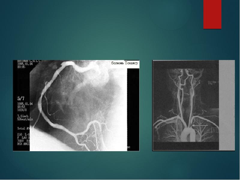
Coronary involvement in TA
Occurs in 10~30%
Often fatal
Classified into 3 types
Type1:stenosis or occlu of coronary ostia
Type2:diffuse or focal coronary arteritis
Type3:coronary aneurysm
Слайд 13
Описание слайда:
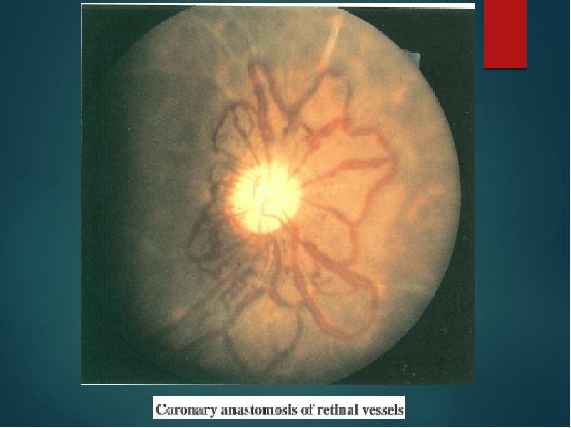
Occular involvement-Amaurosis fugax, pain behind eye, no real visual loss
Hypertensive retinopathy
Слайд 14
Описание слайда:
Слайд 15
Описание слайда:
Слайд 16
Описание слайда:
Ishikawa clinical classification of Takayasu arteritis 1978
Слайд 17
Описание слайда:
Слайд 18
Описание слайда:
Cumulative survival
Cumulative survival
5years -91% (event free survival -74.9%)
10 years -84% (event free survival -64%)
Single mild complication or no complication
5 year event free survival 97%
Single severe or multiple complications
5 year event free survival 59.7%
No deaths in groups I and IIA
19.6% mortality in groups IIB and III (CVA,CCF)
Слайд 19
Описание слайда:
1990
Слайд 20
Описание слайда:
1995
Слайд 21
Описание слайда:
Слайд 22
Описание слайда:
Слайд 23
Описание слайда:
nee
Слайд 24
Описание слайда:
Слайд 25
Описание слайда:
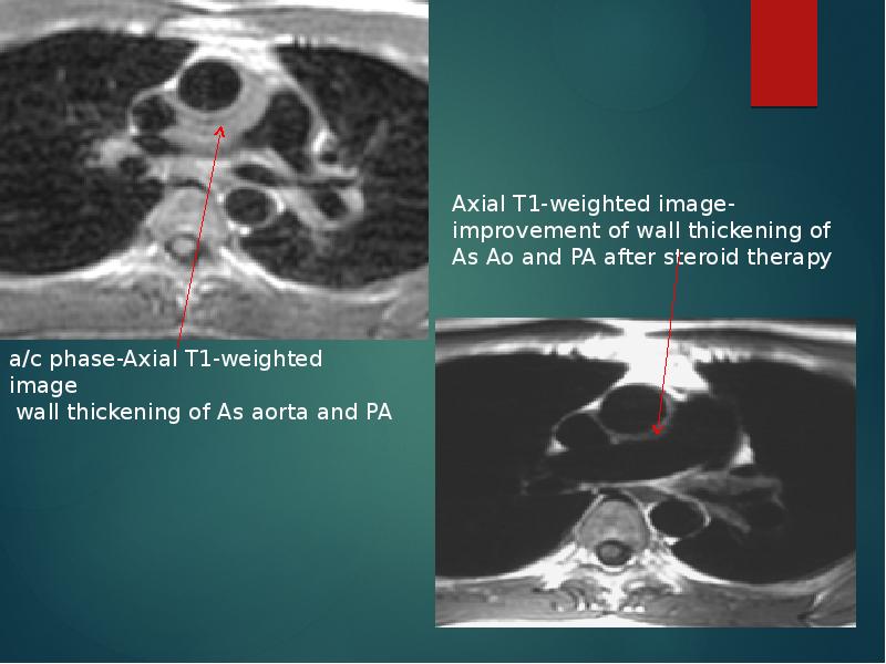
Findings of TA on MRI
mural thrombi
signal alterations within and surrounding inflamed vessels
vascular dilation
thickened aortic valvular cusps
multifocal stenoses
concentric thickening of the aortic wall
Disadvantages
difficulty in visualizing small branch vessels and poor visualization of vascular calcification
may falsely accentuate the degree of vascular stenoses (renal & subclavian)
Слайд 26
Описание слайда:
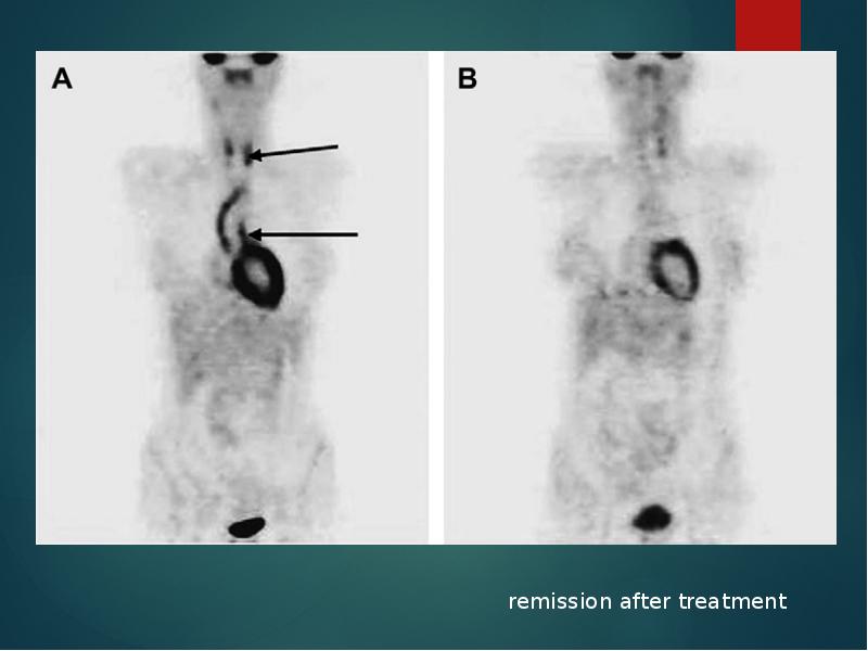
[18F]fluorodeoxyglucose PET for diagnosing
Takayasu’s arteritis
common [18F]FDG uptake pattern TA
early phase - linear and continuous
late phase-patchy rather than continuous ,linear
shown to identify more affected vascular regions than morphologic imaging with MRI
does not provide any information about changes in the wall structure or luminal blood flow
sensitivities of 83% and specificity 100%
( Meller Jet al. Value of F-18 FDG hybrid camera PET and MRI in earlyTakayasu aortitis. Eur Radiol 2003)
Sensitivity of 92%, specificity of 100% and a diagnostic accuracy of 94%
( Webb M et al. The role of 18F-FDG PET in characterising disease activity in Takayasu arteritis. Eur J Nucl Med Imaging 2004
Слайд 27
Описание слайда:
Слайд 28
Описание слайда:
Treatment of TA
・
Слайд 29
Описание слайда:
Medical treatment
Слайд 30
Описание слайда:
Steroids → 50% response
Methotrexate →further 50% respond
25% with active disease will not respond to current treatments
resistant to steroids/ recurrent disease once corticosteroids are tapered
cyclophosphamide (1-2 mg/kg/day),
azathioprine (1-2mg/kg/day), or
methotrexate (0.3 mg/kg/week)
Mycophenolate mofetil/ anti TNF α agents- infliximab
Слайд 31
Описание слайда:
Critical issue is in trying to determine whether or not disease is active
During Rx- regular clinical examination and ESR+ C-RP initially - every few days
CT or MR angio - 3 to 12 months - (active phase of Rx), and annually thereafter
Criteria for active disease
Слайд 32
Описание слайда:
chronic phase- persistent inflammation
steroids should be continued –
<1.0 mg/dL of s.C-RP and 20 mm/h of ESR
Слайд 33
Описание слайда:
Surgical treatment
HTN with critical RAS
Extremity claudication limiting daily activities
Cerebrovascular ischaemia or critical stenoses of ≥3 cerebral vessels
Moderate AR
Cardiac ischaemia with confirmed coronary involvement
Aneurysms
Recommended at quiescent state-avoids compli
(restenosis, anastamotic failure, thrombosis, haemorrhage, & infection)
Слайд 34
Описание слайда:
Surgical techniques
Carry high morbidity & mortality
Steno /aneurysm -anastomotic points
Progressive nature of TA
Diffuse nature of TA
Слайд 35
Описание слайда:
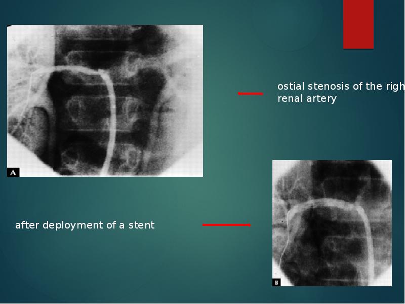
Renal artery involvement
Best treated by PTA
Stent placement following PTA
Ostial lesions
Long segment lesions
Incomplete relief of stenoses
Dissection
Слайд 36
Описание слайда:
Слайд 37
Описание слайда:
Renal PTA - 33 stenoses (20 pts)
Renal PTA - 33 stenoses (20 pts)
Indi-sev HTN,angio 70% stenosis with pr grad 20mm,
nl-ESR
Tech success -28 lesions (85%) clin success-14(82%)
Failures - Coexistent abd Ao disease & tight, prox RAS
Tech diffi - tough, noncompliant stenoses, difficult to cross & resisted repeated, prolonged balloon inflations - backache & ↓SBP during balloon inflation
Follow-up –mean (8/12) -restenosis in 6 (21%)
Renal PTA in TA -tech difficulties; Short-term results - good, Complication rate-acceptable
Слайд 38
Описание слайда:
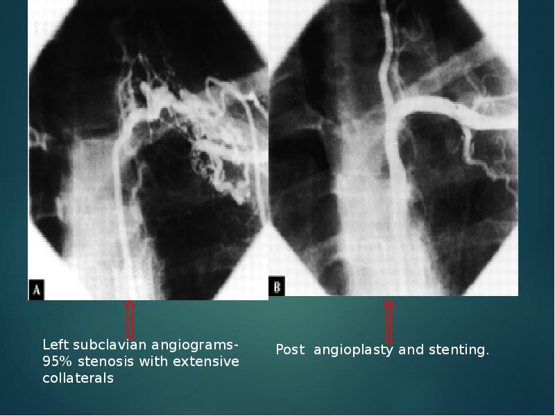
Aortoarteritic lesions
Balloon dilation
safe & reasonably effective
Can be performed repeatedly without any added risks
Balloon dilation diff from atherosclerotic lesions
Minimal intimal involvement –permits easy wiring and balloon crossing
Resistance to dilation – high fibrotic element in the stenotic lesion
restenosis> frequent in TA - diffuse and long stenotic lesions
Слайд 39
Описание слайда:
Слайд 40
Описание слайда:
Joseph s et al, SCT
J Vasc Interv Radiol 1994;5:573–580
Слайд 41
Описание слайда:
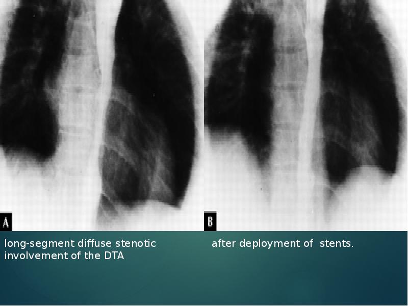
Aortoplasty and Stenting
PTA -desc thoracic and/or abd Ao (TA) stenosis
16 pts (12+4)- HTN/severe b/l- LL claudication
Aortography – stenosis→ DTA-5, abd Ao-10, Both -1
Initial tech & clinical success -100%
patency rate of 67% in a 52-month follow-up
Follow-up (mean 21months)- Restenosis -3
PTA has a definite role in TA management
residual gradient < 20 mm -criterion for successful aortoplasty
long-segment disease, dissection or persistence of a grad > 20 mm Hg after PTBA- aortic stenting
Слайд 42
Описание слайда:
Слайд 43
Описание слайда:
Treatment for cor A occulusion in TA
Surgery (CABG)- often not indicated
・IMA can’t be used often
occlu of Innomi A / Scl A
calcification of aorta
High incidence of restenosis:36%
Angioplasty(PTCA)
・alternative to surgery
Very high incidence of restenosis:78%
DES-effectiveness ?
Слайд 44
Описание слайда:
Percutaneous Management of Aneurysmal Lesions
Aneurysmal dilatation- isolation or together with stenotic lesions
fusiform or saccular
one of the major complications related to the prognosis in TA
Incidence of aneurysm rupture -low
Management - mainly surgical.
Covered stent-grafts may be useful
Скачать презентацию на тему TAKAYASU’S ARTERITIS можно ниже: