Type of Imaging Modalities In Radiology презентация
Содержание
- 2. Medical imaging is the technique and process of creating visual representations of
- 3. As a discipline it is part of biological imaging and incorporates radiology which uses the
- 4. CONVENTIONAL RADIOGRAPHY ■ Images produced through the use of ionizing radiation
- 5. Common uses and disadvantages ■ Common uses for conventional radiography include
- 6. CT or CAT scan
- 7. COMPUTED TOMOGRAPHY ■ CT (or “CAT”) scanners, first introduced in the
- 8. CT or CAT ■ A CT scanner is connected to a
- 9. Traditionally, CT images were viewed mostly in the axial plane. Now,
- 11. CT scans are the cornerstone of cross-sectional imaging and are widely
- 13. ULTRASONOGRAPHY ■ Ultrasound probes utilize acoustic energy above the audible
- 14. Benefits ■ Ultrasound scanners are relatively inexpensive compared with CT and
- 15. ■ Other common uses are evaluation of cystic versus solid breast
- 16. MAGNETIC RESONANCE IMAGING ■ MRI utilizes the potential energy stored in
- 17. ■ MRI is widely used in neurologic imaging and is particularly
- 18. FLUOROSCOPY Fluoroscopy is a modality in which ionizing radiation (x-rays) is
- 19. ■ In interventional radiology, iodinated contrast is selectively injected into blood
- 20. NUCLEAR MEDICINE ■ A radioactive isotope (radioisotope) is an unstable
- 21. Radiopharmaceuticals are combinations of radioisotopes attached to a pharmaceutical that has
- 22. ■ After the radiopharmaceutical is carried to a tissue or organ
- 23. ■ Positron emission tomography (PET) is used to produce three-dimensional images
- 24. ■ PET scanning is most often used in the diagnosis and
- 25. ■ Compared with CT and fluoroscopy, nuclear medicine studies, in general,
- 26. Thank you for your attention !!! This guy partially amputated his
- 27. Literature https://www.google.com https://www.wikipedia.org ^ Jump up to:a b James A.P.; Dasarathy B.V. (2014). "Medical
- 28. Скачать презентацию


 scanners, first introduced in the")


 is")

 is an unstable")


 is used to produce three-dimensional images")


. \"Medical")
Слайды и текст этой презентации
Слайд 1
Описание слайда:
Type of Imaging Modalities
In Radiology
Radiology residents:
Shabelyanov S.
Kuttybaeva A.
Слайд 2
Описание слайда:
Medical imaging is the technique and process of creating visual representations of the interior of a body for clinical analysis and medical intervention, as well as visual representation of the function of some organs or tissues.
Medical imaging is the technique and process of creating visual representations of the interior of a body for clinical analysis and medical intervention, as well as visual representation of the function of some organs or tissues.
Medical imaging seeks to reveal internal structures hidden by the skin and bones, as well as to diagnose and treat disease.
Слайд 3
Описание слайда:
As a discipline it is part of biological imaging and incorporates radiology which uses the imaging technologies of:
As a discipline it is part of biological imaging and incorporates radiology which uses the imaging technologies of:
X-ray,
CT,
ultrasound,
MRI,
and nuclear medicine functional imaging techniques as positron emission tomography (PET) and Single-photon emission computed tomography (SPECT).
Слайд 4
Описание слайда:
CONVENTIONAL RADIOGRAPHY
■ Images produced through the use of ionizing radiation are called conventional radiographs or, more often, plain films.
■ The major advantage of conventional radiographs is that the images are relatively inexpensive to produce, can be obtained almost anywhere by using portable or mobile machines, and are still the most widely obtained imaging studies.
■ They require a source to produce the x-rays (the “x-ray machine”), a method to record the image (a film, cassette, or photosensitive plate), and a way to process the recorded image (using either chemicals or a digital reader).
Слайд 5
Описание слайда:
Common uses and disadvantages
■ Common uses for conventional radiography include the ubiquitous chest x-ray, plain films of the abdomen, and virtually every initial image of the skeletal system to evaluate for fractures or arthritis.
■ The major disadvantages of conventional radiography are the limited range of densities it can demonstrate and that it uses ionizing radiation.
Слайд 6
Описание слайда:
CT or CAT scan
Слайд 7
Описание слайда:
COMPUTED TOMOGRAPHY
■ CT (or “CAT”) scanners, first introduced in the 1970s, brought a quantum leap to medical imaging.
■ Using a gantry with a rotating x-ray beam and multiple detectors in various arrays (which themselves rotate continuously around the patient), along with sophisticated computer algorithms to process the data, a large number of two-dimensional, slicelike images (each of which is millimeters in size) can be formatted in multiple imaging planes.
Слайд 8
Описание слайда:
CT or CAT
■ A CT scanner is connected to a computer that processes the data though various algorithms to produce images of diagnostic quality.
■ A CT image is composed of a matrix of thousands of tiny squares called pixels, each of which is computer-assigned a CT number from −1000 to +1000 measured in Hounsfield units (HUs), named after Sir Godfrey Hounsfield, the man credited with developing the first CT scanner (for which he won the Nobel Prize in Medicine in 1979 with Allan
Cormack).
Слайд 9
Описание слайда:
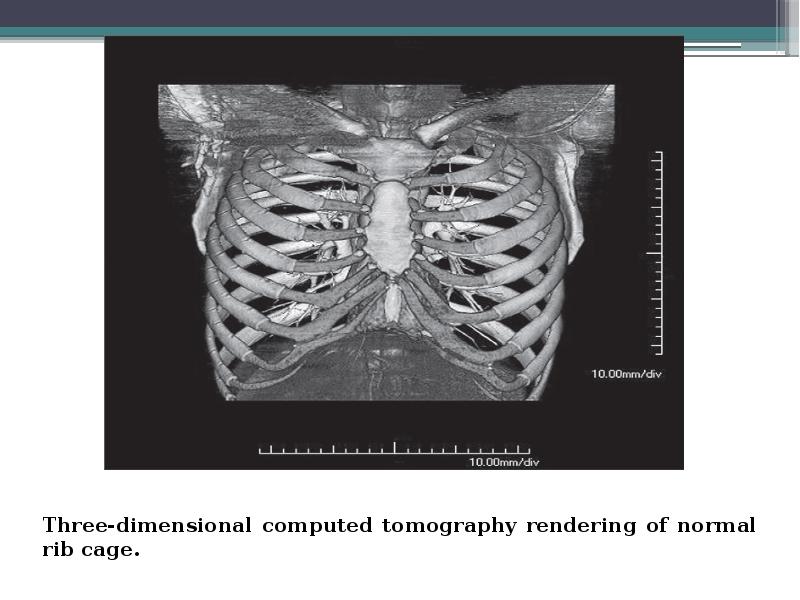
Traditionally, CT images were viewed mostly in the axial plane. Now, because of volumetric acquisition of data, CT scans can be shown in any plane: axial, sagittal, or coronal.
Traditionally, CT images were viewed mostly in the axial plane. Now, because of volumetric acquisition of data, CT scans can be shown in any plane: axial, sagittal, or coronal.
Volumetric data consist of a series of thin sections that can be reassembled for a three-dimensional reconstruction.
Surface and volume rendering in three dimensions can produce CT images of amazing, realistic quality (Next slide).
■ One of the major benefits of CT scanning over conventional radiography is its ability to expand the gray scale, which enables differentiation of many more densities available on conventional radiographs.
Слайд 10
Описание слайда:
Слайд 11
Описание слайда:
CT scans are the cornerstone of cross-sectional imaging and are widely available, although not as yet truly portable.
CT scans are the cornerstone of cross-sectional imaging and are widely available, although not as yet truly portable.
Production of CT images requires an expensive scanner, a space dedicated to its installation, and sophisticated computer processing power. Like conventional x-ray machines, CT scanners utilize ionizing radiation (x-rays) to produce their images.
Слайд 12
Описание слайда:
Слайд 13
Описание слайда:
ULTRASONOGRAPHY
■ Ultrasound probes utilize acoustic energy above the audible frequency of humans to produce images, instead of using x-rays as both conventional radiography and CT scans do.
■ An ultrasound probe or transducer both produces the ultrasonic signal and records it. The signal is processed for its characteristics by an onboard computer. Images are displayed either as static images or in the form of a movie (or “cine”).
Слайд 14
Описание слайда:
Benefits
■ Ultrasound scanners are relatively inexpensive compared with CT and MRI scanners. They are widely available and can be made portable to the point of being handheld.
■ Because ultrasonography utilizes no ionizing radiation, it is particularly useful in obtaining images of children and women of childbearing age and during pregnancy.
■ Ultrasonography is widely used in medical imaging. It is usually the study of first choice in imaging the female pelvis and in pediatric patients, in differentiating cystic versus solid lesions in patients of all ages, in noninvasive vascular imaging, in imaging of the fetus and placenta during pregnancy, and in real-time, image-guided fluid aspiration and biopsy.
Слайд 15
Описание слайда:
■ Other common uses are evaluation of cystic versus solid breast masses, thyroid nodules, and tendons and in assessment of the brain, hips, and spine in newborns. Ultrasonography is used in settings ranging from intraoperative scanning in the surgical suite to the medical tent in the battlefield.
■ Other common uses are evaluation of cystic versus solid breast masses, thyroid nodules, and tendons and in assessment of the brain, hips, and spine in newborns. Ultrasonography is used in settings ranging from intraoperative scanning in the surgical suite to the medical tent in the battlefield.
■ Ultrasonography is generally considered to be a very safe imaging modality that has no known major side effects when used at medically diagnostic levels.
Слайд 16
Описание слайда:
MAGNETIC RESONANCE IMAGING
■ MRI utilizes the potential energy stored in the body’s hydrogen atoms. The atoms are manipulated by very strong magnetic fields and radiofrequency pulses to produce enough localizing and tissue-specific energy to allow highly sophisticated computer programs to generate two- and three dimensional images.
■ However, they utilize no ionizing radiation and produce much higher contrast between different types of soft tissues than is possible with CT.
Слайд 17
Описание слайда:
■ MRI is widely used in neurologic imaging and is particularly sensitive in imaging soft tissues such as the muscles, tendons, and ligaments.
■ MRI is widely used in neurologic imaging and is particularly sensitive in imaging soft tissues such as the muscles, tendons, and ligaments.
■Сontraindications of MRI:
Cardiac pacemaker or metal valve.
Brain aneurysm and aortic clips
Endoprosthesis
Metal objects:
Within the eye;
Near the spinal cord;
Cochlear implants;
Insulin pumps;
Neurostimulators.
Claustrophobia
Слайд 18
Описание слайда:
FLUOROSCOPY
Fluoroscopy is a modality in which ionizing radiation (x-rays) is used in performing real-time visualization of the body in a way that allows for evaluation of the motion of body parts, real-time positioning changes of bones and joints, and the location and path of externally administered barium or iodine contrast agents through the gastrointestinal and genitourinary tracts and blood vessels. Images can be viewed as they are acquired on video screens and captured as either a series of static images or moving (video) images.
Слайд 19
Описание слайда:
■ In interventional radiology, iodinated contrast is selectively injected into blood vessels or other ducts that can be imaged fluoroscopically to demonstrate normal anatomy, pathology, or the position of catheters or other devices
■ In interventional radiology, iodinated contrast is selectively injected into blood vessels or other ducts that can be imaged fluoroscopically to demonstrate normal anatomy, pathology, or the position of catheters or other devices
■ Radiation doses in fluoroscopy can be substantially higher than those used in conventional radiography because so many images are acquired for every minute of fluoroscopy time. Therefore the dose is reduced by using the shortest possible fluoroscopy time to obtain diagnostic images.
Слайд 20
Описание слайда:
NUCLEAR MEDICINE
■ A radioactive isotope (radioisotope) is an unstable form of an element that emits radiation from its nucleus as it decays. Eventually, the end product is a stable, nonradioactive isotope of another element.
■ Radioisotopes can be produced artificially (most frequently by neutron enrichment in a nuclear reactor or in a cyclotron) or may occur naturally. Naturally occurring radioisotopes include uranium and thorium. The vast majority of radioisotopes used in medicine are produced artificially.
Слайд 21
Описание слайда:
Radiopharmaceuticals are combinations of radioisotopes attached to a pharmaceutical that has binding properties that allow it to concentrate in certain body tissues, such as the lungs, thyroid, or bones. Radioisotopes used in clinical nuclear medicine are also referred to as radionuclides, radiotracers, or, sometimes, simply tracers.
Radiopharmaceuticals are combinations of radioisotopes attached to a pharmaceutical that has binding properties that allow it to concentrate in certain body tissues, such as the lungs, thyroid, or bones. Radioisotopes used in clinical nuclear medicine are also referred to as radionuclides, radiotracers, or, sometimes, simply tracers.
■ Various body organs have a specific affinity for, or absorption of, different biologically active chemicals. For example, the thyroid takes up iodine; the brain utilizes glucose; bones utilize phosphates; and particles of a certain size can be trapped in the lung capillaries.
Слайд 22
Описание слайда:
■ After the radiopharmaceutical is carried to a tissue or organ in the body, usually via the bloodstream, its radioactive emissionsn allow it to be measured and imaged using a detection device called a gamma camera.
■ After the radiopharmaceutical is carried to a tissue or organ in the body, usually via the bloodstream, its radioactive emissionsn allow it to be measured and imaged using a detection device called a gamma camera.
Слайд 23
Описание слайда:
■ Positron emission tomography (PET) is used to produce three-dimensional images that depict the body’s biochemical and metabolic processes at a molecular level. It is performed using a positron (positive electron)-producing radioisotope attached to a targeting pharmaceutical.
■ Positron emission tomography (PET) is used to produce three-dimensional images that depict the body’s biochemical and metabolic processes at a molecular level. It is performed using a positron (positive electron)-producing radioisotope attached to a targeting pharmaceutical.
Слайд 24
Описание слайда:
■ PET scanning is most often used in the diagnosis and treatment follow-up of cancer. It is frequently used to locate hidden metastases from a known tumor or to detect recurrence. Oncologic PET scans make up about 90% of the clinical use of PET.
■ PET scanning is most often used in the diagnosis and treatment follow-up of cancer. It is frequently used to locate hidden metastases from a known tumor or to detect recurrence. Oncologic PET scans make up about 90% of the clinical use of PET.
■ Unlike other modalities that use ionizing radiation, the patient can briefly be the source of radiation exposure to others (e.g., technologists) in nuclear medicine studies. To limit exposure to others, the principles of decreasing the time in close proximity to the patient, increasing the distance from the source, and appropriate shielding are used.
Слайд 25
Описание слайда:
■ Compared with CT and fluoroscopy, nuclear medicine studies, in general, produce less patient exposure.
■ Compared with CT and fluoroscopy, nuclear medicine studies, in general, produce less patient exposure.
The types of scans that deliver the highest dose relative to other nuclear scans are cardiac studies and PET examinations.
Слайд 26
Описание слайда:
Thank you for your attention !!!
This guy partially amputated his finger on a circular saw. "I stopped paying attention for one second!" he said.
Слайд 27
Описание слайда:
Literature
https://www.google.com
https://www.wikipedia.org
^ Jump up to:a b James A.P.; Dasarathy B.V. (2014). "Medical Image Fusion: A survey of state of the art". Information Fusion. 19: 4–19. arXiv:1401.0166. doi:10.1016/j.inffus.2013.12.002.
Скачать презентацию на тему Type of Imaging Modalities In Radiology можно ниже: